Fluorescence Diffraction Tomography using Explicit Neural Fields
Publication
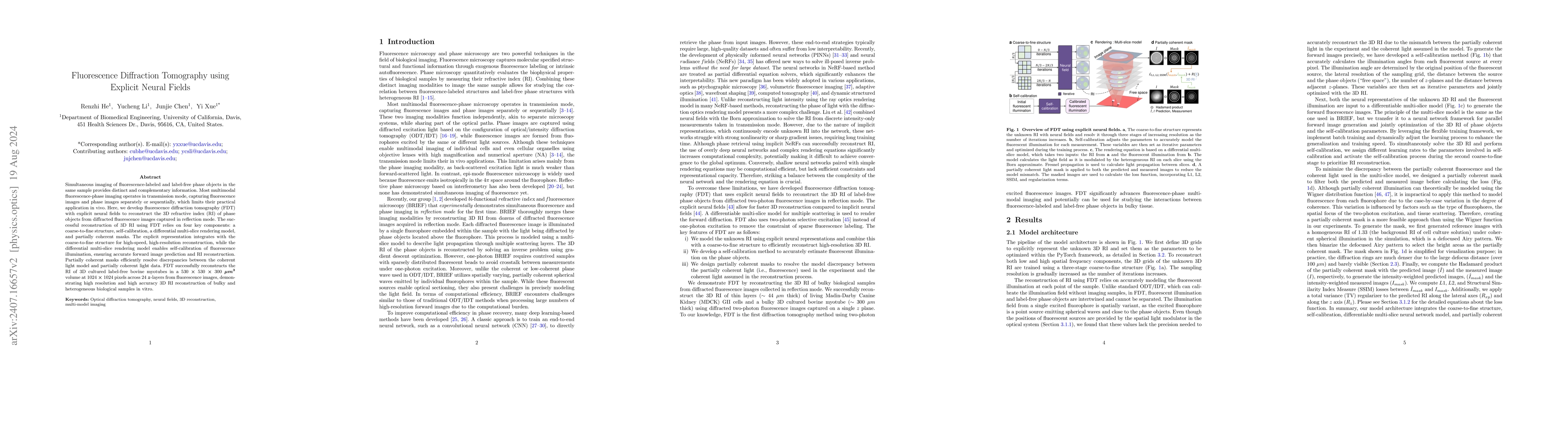
Metrics
AI Quick Summary
This paper introduces fluorescence diffraction tomography (FDT) with explicit neural fields to reconstruct the 3D refractive index of phase objects using diffracted fluorescence images in reflection mode. FDT integrates a coarse-to-fine structure, self-calibration, differential multi-slice rendering, and partially coherent masks for high-speed, high-resolution 3D RI reconstruction, successfully demonstrating its application on cultured bovine myotubes.
Paper Preview
Abstract
Simultaneous imaging of fluorescence-labeled and label-free phase objects in the same sample provides distinct and complementary information. Most multimodal fluorescence-phase imaging operates in transmission mode, capturing fluorescence images and phase images separately or sequentially, which limits their practical application in vivo. Here, we develop fluorescence diffraction tomography (FDT) with explicit neural fields to reconstruct the 3D refractive index (RI) of phase objects from diffracted fluorescence images captured in reflection mode. The successful reconstruction of 3D RI using FDT relies on four key components: a coarse-to-fine structure, self-calibration, a differential multi-slice rendering model, and partially coherent masks. The explicit representation integrates with the coarse-to-fine structure for high-speed, high-resolution reconstruction, while the differential multi-slice rendering model enables self-calibration of fluorescence illumination, ensuring accurate forward image prediction and RI reconstruction. Partially coherent masks efficiently resolve discrepancies between the coherent light model and partially coherent light data. FDT successfully reconstructs the RI of 3D cultured label-free bovine myotubes in a 530 $\times$ 530 $\times$ 300 $\mu m^3$ volume at 1024 $\times$ 1024 pixels across 24 $z$-layers from fluorescence images, demonstrating high resolution and high accuracy 3D RI reconstruction of bulky and heterogeneous biological samples in vitro.
AI Key Findings
Get AI-generated insights about this paper's methodology, results, significance, and more — seven facets brought into focus.
Impact
Authors
PDF Preview
Citation Network
Current paper (gray), citations (green), references (blue)
Display is limited for performance on very large graphs.
Discussion 0