Authors
Publication
Metrics
Quick Actions
Quick Answers
What methodology did the authors use?
The research proposes a hybrid deep learning architecture combining convolutional neural networks (CNNs) and transformers for medical image segmentation, specifically for polyp and breast ultrasound image analysis. It evaluates the model using benchmark datasets and compares performance against existing state-of-the-art methods. More in Methodology →
What are the key results?
The proposed TBConvL-Net architecture achieves state-of-the-art performance on polyp segmentation tasks — The model demonstrates competitive results compared to existing methods like U-Net and Swin-Unet More in Key Results →
Why is this work significant?
This research advances medical image analysis by providing a more effective segmentation solution that could improve diagnostic accuracy and efficiency in clinical settings More in Significance →
What are the main limitations?
The model's performance may degrade with highly variant ultrasound imaging conditions — The approach requires substantial computational resources for training More in Limitations →
Abstract
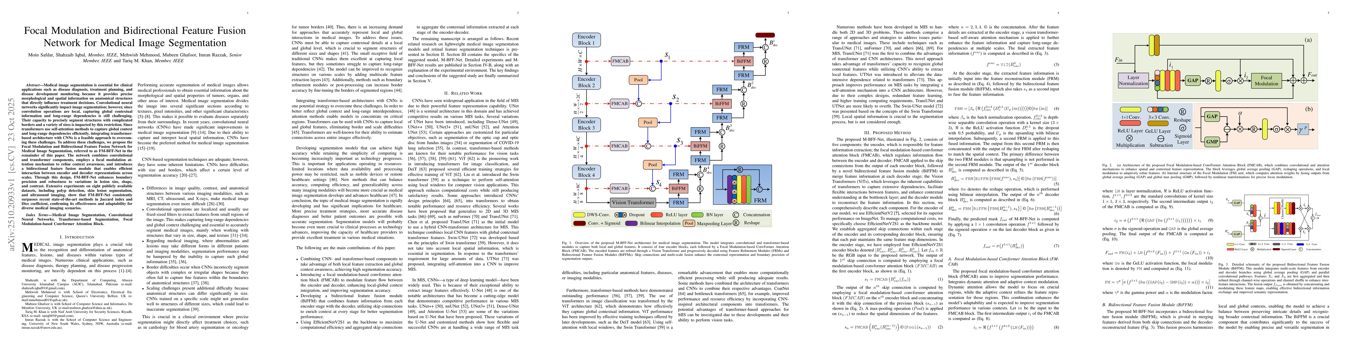
Medical image segmentation is essential for clinical applications such as disease diagnosis, treatment planning, and disease development monitoring because it provides precise morphological and spatial information on anatomical structures that directly influence treatment decisions. Convolutional neural networks significantly impact image segmentation; however, since convolution operations are local, capturing global contextual information and long-range dependencies is still challenging. Their capacity to precisely segment structures with complicated borders and a variety of sizes is impacted by this restriction. Since transformers use self-attention methods to capture global context and long-range dependencies efficiently, integrating transformer-based architecture with CNNs is a feasible approach to overcoming these challenges. To address these challenges, we propose the Focal Modulation and Bidirectional Feature Fusion Network for Medical Image Segmentation, referred to as FM-BFF-Net in the remainder of this paper. The network combines convolutional and transformer components, employs a focal modulation attention mechanism to refine context awareness, and introduces a bidirectional feature fusion module that enables efficient interaction between encoder and decoder representations across scales. Through this design, FM-BFF-Net enhances boundary precision and robustness to variations in lesion size, shape, and contrast. Extensive experiments on eight publicly available datasets, including polyp detection, skin lesion segmentation, and ultrasound imaging, show that FM-BFF-Net consistently surpasses recent state-of-the-art methods in Jaccard index and Dice coefficient, confirming its effectiveness and adaptability for diverse medical imaging scenarios.
AI Key Findings
Generated Nov 15, 2025
Methodology — What approach did the authors take?
The research proposes a hybrid deep learning architecture combining convolutional neural networks (CNNs) and transformers for medical image segmentation, specifically for polyp and breast ultrasound image analysis. It evaluates the model using benchmark datasets and compares performance against existing state-of-the-art methods.
Key Results — What are the main findings?
- The proposed TBConvL-Net architecture achieves state-of-the-art performance on polyp segmentation tasks
- The model demonstrates competitive results compared to existing methods like U-Net and Swin-Unet
- The approach shows promising potential for breast ultrasound image analysis
Significance — Why does this research matter?
This research advances medical image analysis by providing a more effective segmentation solution that could improve diagnostic accuracy and efficiency in clinical settings
Technical Contribution — What is the technical contribution?
Development of a hybrid CNN-transformer architecture with specialized attention mechanisms for medical image segmentation
Novelty — What is new about this work?
Integration of transformer-based attention mechanisms with CNNs in a novel architecture specifically optimized for medical image segmentation tasks
Limitations — What are the limitations of this study?
- The model's performance may degrade with highly variant ultrasound imaging conditions
- The approach requires substantial computational resources for training
Future Work — What did the authors propose for future work?
- Exploring domain adaptation techniques to improve performance across different imaging modalities
- Investigating model compression techniques to enable deployment on resource-constrained devices
How to Cite This Paper
@article{razzak2025focal,
title = {Focal Modulation and Bidirectional Feature Fusion Network for Medical
Image Segmentation},
author = {Razzak, Imran and Mehmood, Mehwish and Safdar, Moin and others},
year = {2025},
eprint = {2510.20933},
archivePrefix = {arXiv},
primaryClass = {cs.CV},
}
Razzak, I., Mehmood, M., Safdar, M., Ghafoor, M., Iqbal, S., & Khan, T. (2025). Focal Modulation and Bidirectional Feature Fusion Network for Medical
Image Segmentation. arXiv. https://arxiv.org/abs/2510.20933
Razzak, Imran, et al. "Focal Modulation and Bidirectional Feature Fusion Network for Medical
Image Segmentation." arXiv, 2025, arxiv.org/abs/2510.20933.
PDF Preview
Similar Papers
Found 4 papersFocal-UNet: UNet-like Focal Modulation for Medical Image Segmentation
MohammadReza Naderi, MohammadHossein Givkashi, Fatemeh Piri et al.
BSAFusion: A Bidirectional Stepwise Feature Alignment Network for Unaligned Medical Image Fusion
Huafeng Li, Dayong Su, Qing Cai et al.
DCFFSNet: Deep Connectivity Feature Fusion Separation Network for Medical Image Segmentation
Xun Ye, Ruixiang Tang, Mingda Zhang et al.
Boundary feature fusion network for tooth image segmentation
Dongping Zhang, Zheng Li, Fangao Zeng et al.
Comments (0)