ArxivLens

Quick Summary:

This paper presents a novel force sensing guided segmentation method to improve artery-vein differentiation in ultrasound images by leveraging vascular deformability. The proposed framework uses key frames identified by force magnitude to enhance segmentation accuracy and integrates a multimodal dataset, Mus-V, for training and evaluation.

arXiv Id: 2407.21394
Date Published: 2024-07-31
Date Updated: 2024-07-31

Categories:

Key Terms (TF-IDF):

ultrasound (0.359)
force (0.267)
segmentation (0.261)
guided (0.191)
frames (0.188)
sensing (0.167)
dataset (0.164)
images (0.164)
integrated (0.157)
magnitude (0.142)
key (0.125)
morphological (0.121)
comprises (0.120)
similarities (0.118)
networks (0.118)
significant (0.118)
robotics (0.117)
current (0.117)
assigned (0.116)
multimodal (0.115)

Summary:

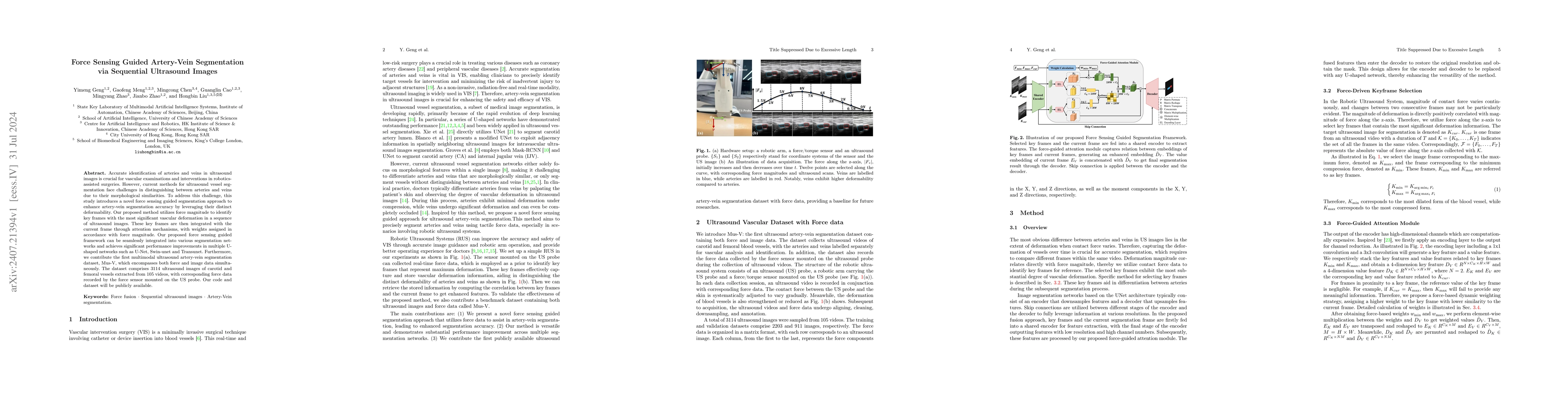
Accurate identification of arteries and veins in ultrasound images is crucial for vascular examinations and interventions in robotics-assisted surgeries. However, current methods for ultrasound vessel segmentation face challenges in distinguishing between arteries and veins due to their morphological similarities. To address this challenge, this study introduces a novel force sensing guided segmentation approach to enhance artery-vein segmentation accuracy by leveraging their distinct deformability. Our proposed method utilizes force magnitude to identify key frames with the most significant vascular deformation in a sequence of ultrasound images. These key frames are then integrated with the current frame through attention mechanisms, with weights assigned in accordance with force magnitude. Our proposed force sensing guided framework can be seamlessly integrated into various segmentation networks and achieves significant performance improvements in multiple U-shaped networks such as U-Net, Swin-unet and Transunet. Furthermore, we contribute the first multimodal ultrasound artery-vein segmentation dataset, Mus-V, which encompasses both force and image data simultaneously. The dataset comprises 3114 ultrasound images of carotid and femoral vessels extracted from 105 videos, with corresponding force data recorded by the force sensor mounted on the US probe. Our code and dataset will be publicly available.

Citation & Reference Network Visualization

This graph shows how this paper is connected to other research. The current paper is shown in dark gray, papers that cite it are in green, and papers it references are in blue. You can:

Current Paper
Papers citing this work (Citations)
Papers cited by this work (References)

• Hover over any node to see the paper title

• Click a node to view that paper's details

• Drag nodes to rearrange the graph

• Use the zoom controls (bottom right) to zoom in/out

Citations and References

No citations found for this paper.

Similar Papers

CAVE: Cerebral Artery-Vein Segmentation in Digital Subtraction Angiography

Ruisheng Su, Yuan Chen, P. Matthijs van der Sluijs, Sandra Cornelissen, Ruben van den Broek, Wim H. van Zwam, Aad van der Lugt, Wiro Niessen, Danny Ruijters, Theo van Walsum

Published: 2024-05-02

Self-adaptive vision-language model for 3D segmentation of pulmonary artery and vein

Lijun Zhang, Yanda Meng, Haochen Zhao, Dan Wang, Tao Zhou, Yuan Li, Xiaotong Guo, Deqian Yang, Zhilin Sui

Published: 2025-01-07

Comments (0)

This website uses cookies to ensure you get the best experience. Learn more