Formation of protein-mediated tubes is governed by a snapthrough transition
Publication
Metrics
AI Quick Summary
This paper presents a mathematical model to describe the formation of protein-mediated plasma membrane tubes, revealing that their length undergoes a snapthrough transition influenced by membrane tension and protein-induced curvature. The study highlights the implications for cellular processes involving BAR-domain proteins.
Paper Preview
Abstract
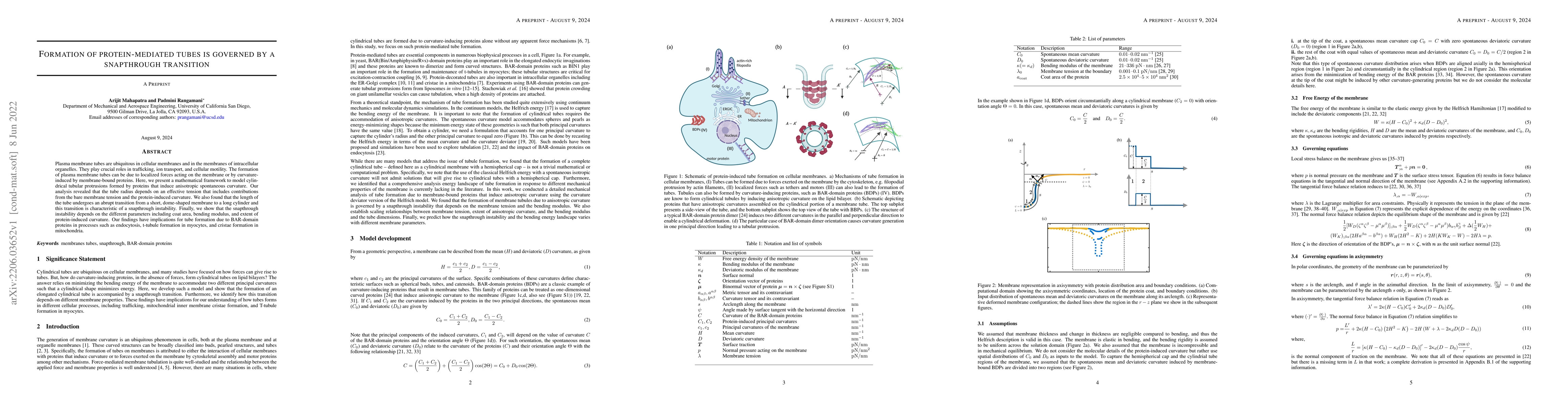
Plasma membrane tubes are ubiquitous in cellular membranes and in the membranes of intracellular organelles. They play crucial roles in trafficking, ion transport, and cellular motility. The formation of plasma membrane tubes can be due to localized forces acting on the membrane or by curvature-induced by membrane-bound proteins. Here, we present a mathematical framework to model cylindrical tubular protrusions formed by proteins that induce anisotropic spontaneous curvature. Our analysis revealed that the tube radius depends on an effective tension that includes contributions from the bare membrane tension and the protein-induced curvature. We also found that the length of the tube undergoes an abrupt transition from a short, dome-shaped membrane to a long cylinder and this transition is characteristic of a snapthrough instability. Finally, we show that the snapthrough instability depends on the different parameters including coat area, bending modulus, and extent of protein-induced curvature. Our findings have implications for tube formation due to BAR-domain proteins in processes such as endocytosis, t-tubule formation in myocytes, and cristae formation in mitochondria.
AI Key Findings
Get AI-generated insights about this paper's methodology, results, significance, and more — seven facets brought into focus.
Impact
Paper Details
Authors
PDF Preview
Key Terms
Citation Network
Current paper (gray), citations (green), references (blue)
Display is limited for performance on very large graphs.
Discussion 0