Formula-Driven Data Augmentation and Partial Retinal Layer Copying for Retinal Layer Segmentation
Publication
Metrics
AI Quick Summary
This research proposes formula-driven data augmentation (FDDA) and partial retinal layer copying (PRLC) to enhance retinal layer segmentation in OCT images without requiring retinal flattening. These methods improve segmentation accuracy for varied retinal structures, validated through experiments on multiple datasets.
Paper Preview
Abstract
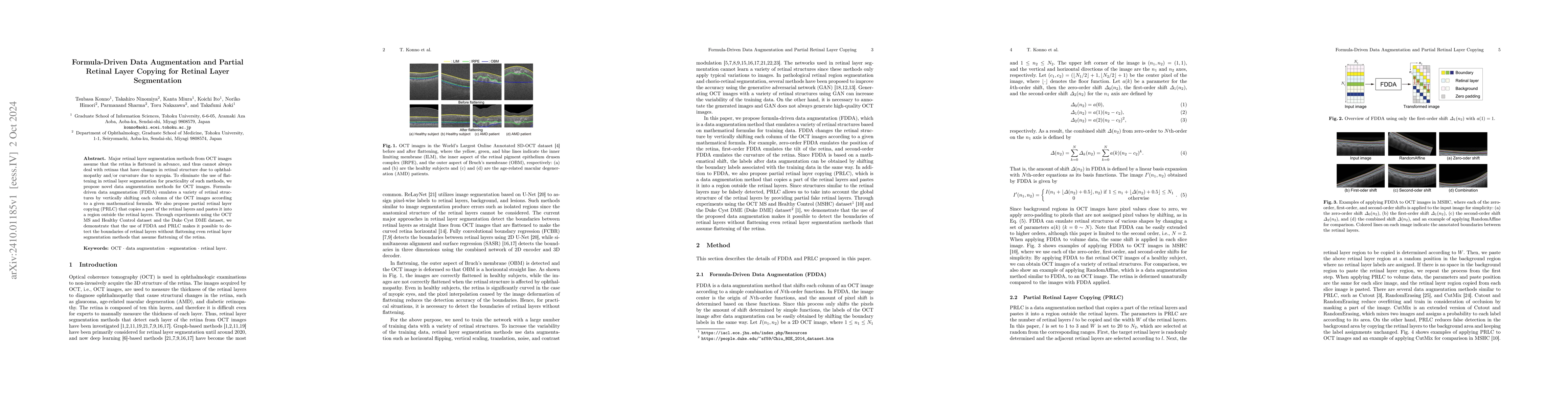
Major retinal layer segmentation methods from OCT images assume that the retina is flattened in advance, and thus cannot always deal with retinas that have changes in retinal structure due to ophthalmopathy and/or curvature due to myopia. To eliminate the use of flattening in retinal layer segmentation for practicality of such methods, we propose novel data augmentation methods for OCT images. Formula-driven data augmentation (FDDA) emulates a variety of retinal structures by vertically shifting each column of the OCT images according to a given mathematical formula. We also propose partial retinal layer copying (PRLC) that copies a part of the retinal layers and pastes it into a region outside the retinal layers. Through experiments using the OCT MS and Healthy Control dataset and the Duke Cyst DME dataset, we demonstrate that the use of FDDA and PRLC makes it possible to detect the boundaries of retinal layers without flattening even retinal layer segmentation methods that assume flattening of the retina.
AI Key Findings
Get AI-generated insights about this paper's methodology, results, significance, and more — seven facets brought into focus.
Impact
Paper Details
Authors
PDF Preview
Citation Network
Current paper (gray), citations (green), references (blue)
Display is limited for performance on very large graphs.
Discussion 0