Quick Summary:

This paper proposes a foundation model for whole-heart segmentation using self-supervised learning and a student-teacher architecture, pre-trained on large unlabeled CT and MRI datasets. The model leverages xLSTM to capture spatial dependencies and achieves high generalization across modalities, reducing reliance on extensive labeled data.

Summary

Whole-heart segmentation from CT and MRI scans is crucial for cardiovascular disease analysis, yet existing methods struggle with modality-specific biases 
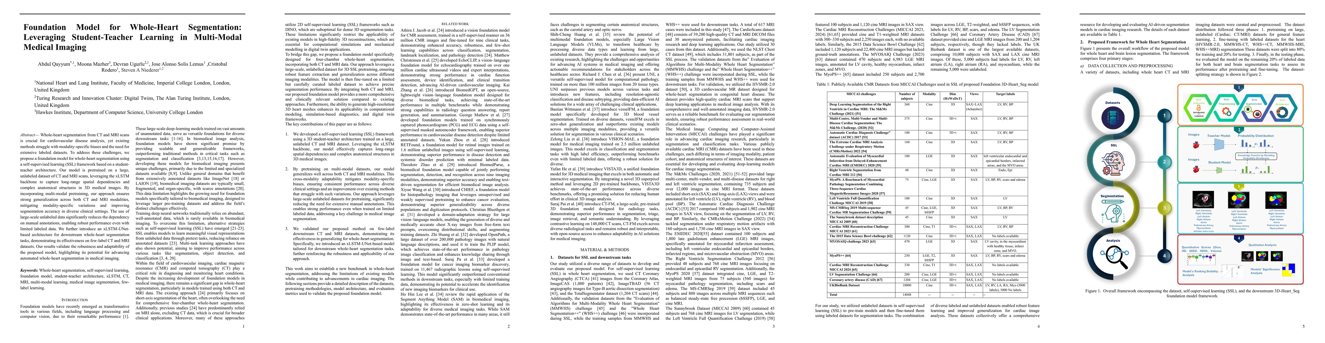
and the need for extensive labeled datasets. To address these challenges, we propose a foundation model for whole-heart segmentation using a self-supervised learning (SSL) framework based on a student-teacher architecture. Our model is pretrained on a large, unlabeled dataset of CT and MRI scans, leveraging the xLSTM backbone to capture long-range spatial dependencies and complex anatomical structures in 3D medical images. By incorporating multi-modal pretraining, our approach ensures strong generalization across both CT and MRI modalities, mitigating modality-specific variations and improving segmentation accuracy in diverse clinical settings. The use of large-scale unlabeled data significantly reduces the dependency on manual annotations, enabling robust performance even with limited labeled data. We further introduce an xLSTM-UNet-based architecture for downstream whole-heart segmentation tasks, demonstrating its effectiveness on few-label CT and MRI datasets. Our results validate the robustness and adaptability of the proposed model, highlighting its potential for advancing automated whole-heart segmentation in medical imaging.

AI Key Findings

Generated Jun 10, 2025

Methodology

The research proposes a foundation model for whole-heart segmentation using a self-supervised learning (SSL) framework based on a student-teacher architecture, pretrained on a large, unlabeled dataset of CT and MRI scans, leveraging the xLSTM backbone to capture long-range spatial dependencies and complex anatomical structures in 3D medical images.

Key Results

  • The proposed model outperforms existing state-of-the-art (SOTA) segmentation models across all datasets, achieving the highest Dice coefficients and significantly lower Hausdorff Distance 95% (HD95) values.
  • The model demonstrates robust performance even with limited labeled data, reducing dependency on manual annotations.
  • Multi-modal pretraining ensures strong generalization across both CT and MRI modalities, mitigating modality-specific variations.

Significance

This research is significant as it addresses the challenges of modality-specific biases and the need for extensive labeled datasets in whole-heart segmentation, offering a more efficient and scalable solution for clinical applications.

Technical Contribution

The introduction of a self-supervised learning framework with a student-teacher architecture and xLSTM backbone for capturing complex 3D anatomical structures in medical imaging.

Novelty

This work stands out by combining self-supervised learning with a student-teacher approach and multi-modal pretraining, enabling robust and accurate whole-heart segmentation across diverse clinical settings with minimal labeled data.

Limitations

  • The model's performance heavily relies on the quality and diversity of the unlabeled dataset used for pretraining.
  • While the model shows robustness, it may still face challenges with highly atypical or rare cardiac anatomies not well-represented in the training data.

Future Work

  • Investigate the model's performance on other cardiovascular structures and diseases beyond whole-heart segmentation.
  • Explore the integration of domain adaptation techniques to further improve the model's generalization to new, unseen datasets.

Paper Details

PDF Preview

Download PDF

Key Terms

No key terms available for this paper.

No citations found for this paper.

Comments (0)

This website uses cookies to ensure you get the best experience. Learn more