Publication
Metrics
AI Quick Summary
Freecyto is a web-based application for interactive flow cytometry data analysis using a weighted k-means clustering algorithm to quantize large datasets, enabling visualization and standard FCM features. It maintains data accuracy comparable to conventional software while addressing browser limitations.
Paper Preview
Abstract
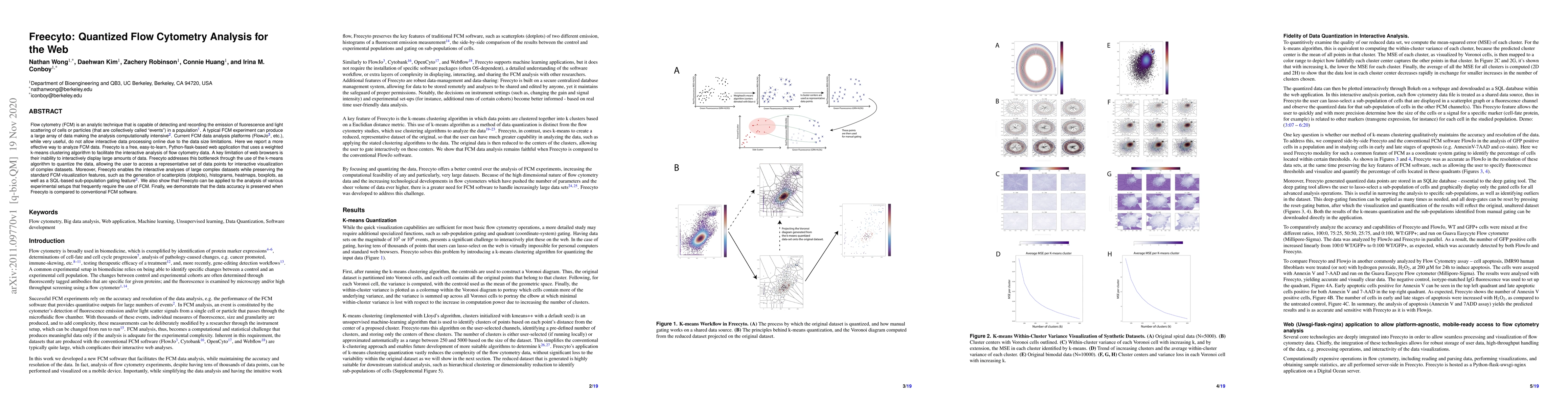
Flow cytometry (FCM) is an analytic technique that is capable of detecting and recording the emission of fluorescence and light scattering of cells or particles (that are collectively called "events") in a population. A typical FCM experiment can produce a large array of data making the analysis computationally intensive. Current FCM data analysis platforms (FlowJo, etc.), while very useful, do not allow interactive data processing online due to the data size limitations. Here we report a more effective way to analyze FCM data. Freecyto is a free, easy-to-learn, Python-flask-based web application that uses a weighted k-means clustering algorithm to facilitate the interactive analysis of flow cytometry data. A key limitation of web browsers is their inability to interactively display large amounts of data. Freecyto addresses this bottleneck through the use of the k-means algorithm to quantize the data, allowing the user to access a representative set of data points for interactive visualization of complex datasets. Moreover, Freecyto enables the interactive analyses of large complex datasets while preserving the standard FCM visualization features, such as the generation of scatterplots (dotplots), histograms, heatmaps, boxplots, as well as a SQL-based sub-population gating feature. We also show that Freecyto can be applied to the analysis of various experimental setups that frequently require the use of FCM. Finally, we demonstrate that the data accuracy is preserved when Freecyto is compared to conventional FCM software.
AI Key Findings
Get AI-generated insights about this paper's methodology, results, significance, and more — seven facets brought into focus.
Impact
Paper Details
Authors
PDF Preview
Key Terms
Citation Network
Current paper (gray), citations (green), references (blue)
Display is limited for performance on very large graphs.
Discussion 0