Summary

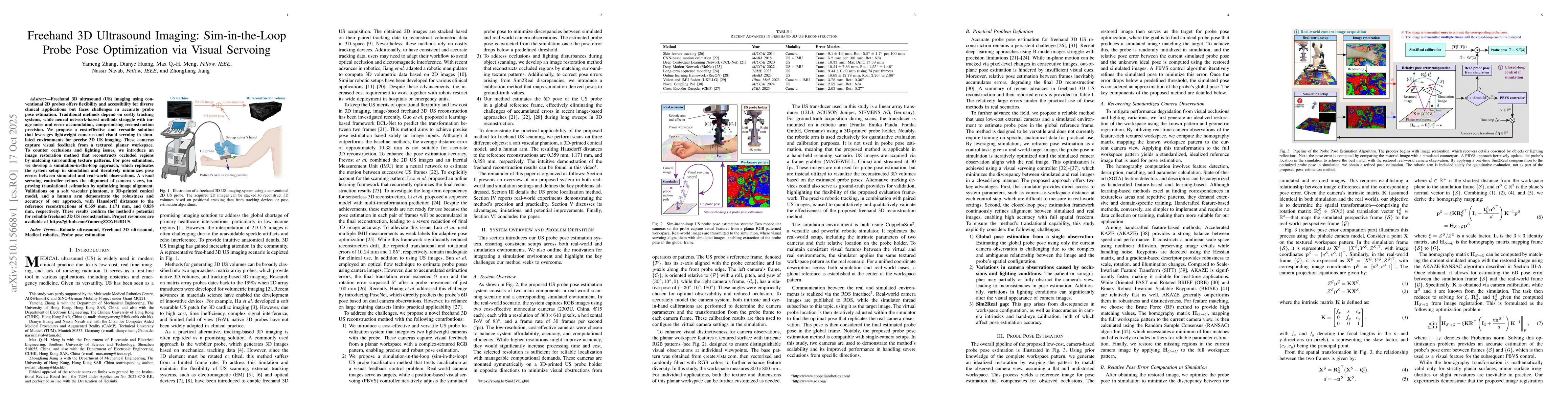
Freehand 3D ultrasound (US) imaging using conventional 2D probes offers flexibility and accessibility for diverse clinical applications but faces challenges in accurate probe pose estimation. Traditional methods depend on costly tracking systems, while neural network-based methods struggle with image noise and error accumulation, compromising reconstruction precision. We propose a cost-effective and versatile solution that leverages lightweight cameras and visual servoing in simulated environments for precise 3D US imaging. These cameras capture visual feedback from a textured planar workspace. To counter occlusions and lighting issues, we introduce an image restoration method that reconstructs occluded regions by matching surrounding texture patterns. For pose estimation, we develop a simulation-in-the-loop approach, which replicates the system setup in simulation and iteratively minimizes pose errors between simulated and real-world observations. A visual servoing controller refines the alignment of camera views, improving translational estimation by optimizing image alignment. Validations on a soft vascular phantom, a 3D-printed conical model, and a human arm demonstrate the robustness and accuracy of our approach, with Hausdorff distances to the reference reconstructions of 0.359 mm, 1.171 mm, and 0.858 mm, respectively. These results confirm the method's potential for reliable freehand 3D US reconstruction.

AI Key Findings

Generated Oct 21, 2025

Methodology

The research proposes a cost-effective solution for freehand 3D ultrasound reconstruction using two lightweight monocular cameras mounted on the ultrasound probe. It employs a simulated-in-the-loop framework with a PBVS controller to iteratively minimize pose errors between simulated and real-world observations. An image restoration technique is introduced to recover details obscured by occlusions or lighting variations.

Key Results

  • The method achieves high accuracy in both positional and rotational estimations
  • Freehand US scans on a soft vascular phantom, 3D-printed conical model, and human arm yielded Hausdorff distances of 0.359mm, 1.171mm, and 0.858mm respectively
  • The system provides precise, robust, and practical solutions for US probe pose estimation and freehand 3D US reconstruction

Significance

This research addresses critical challenges in freehand ultrasound imaging by providing accurate probe pose estimation without relying on noisy ultrasound images. It enables clearer anatomical visualization for more reliable diagnosis, reduces dependence on expensive tracking equipment, and supports medical training and skill assessment.

Technical Contribution

Development of a simulated-in-the-loop framework with PBVS controller for accurate pose estimation and image restoration technique for occlusion/lighting variation recovery

Novelty

Integration of visual feedback from a textured planar workspace with a closed-loop image matching mechanism, combined with image restoration techniques, provides a novel approach to probe pose estimation in freehand ultrasound applications

Limitations

  • The system cannot tolerate fully occluded camera FoV
  • Real-time pose estimation has not yet been achieved

Future Work

  • Incorporate a laser tracker for more precise localization assessment
  • Explore compatibility with various probe types and advanced camera configurations

Paper Details

PDF Preview

Download PDF

Comments (0)

This website uses cookies to ensure you get the best experience. Learn more