Full-Field Interferometric Imaging of Propagating Action Potentials
Publication
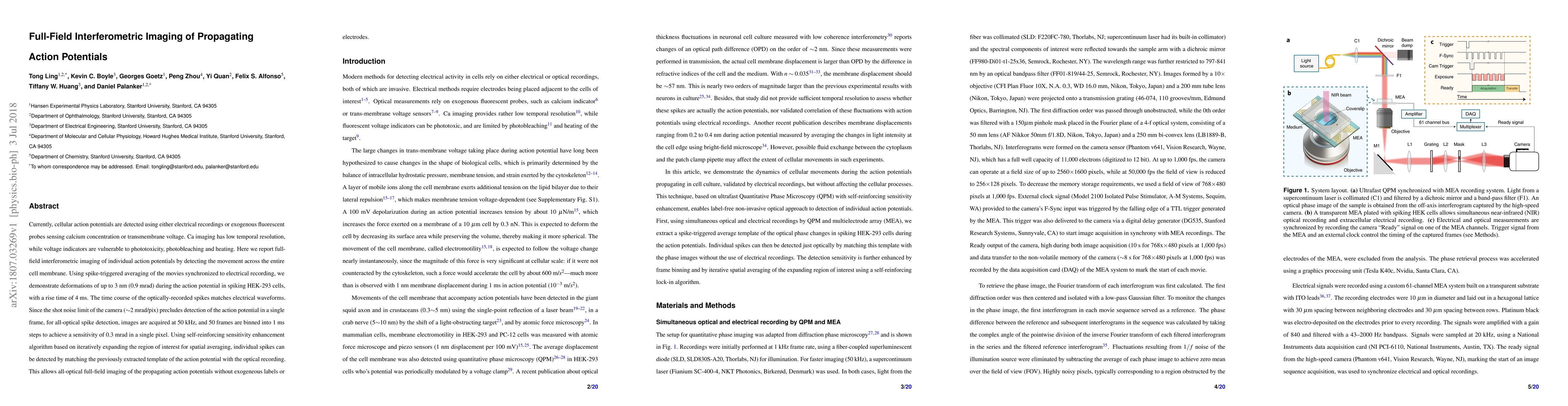
Metrics
AI Quick Summary
This paper presents a novel full-field interferometric imaging technique for detecting individual action potentials across cell membranes without using electrical recordings or fluorescent probes. The method achieves high sensitivity by binning 50 frames per millisecond and employing a self-reinforcing algorithm to match the action potential template, enabling all-optical detection of propagating spikes in HEK-293 cells.
Paper Preview
Abstract
Currently, cellular action potentials are detected using either electrical recordings or exogenous fluorescent probes sensing calcium concentration or transmembrane voltage. Ca imaging has low temporal resolution, while voltage indicators are vulnerable to phototoxicity, photobleaching and heating. Here we report full-field interferometric imaging of individual action potentials by detecting the movement across the entire cell membrane. Using spike-triggered averaging of the movies synchronized to electrical recording, we demonstrate deformations of up to 3 nm (0.9 mrad) during the action potential in spiking HEK-293 cells, with a rise time of 4 ms. The time course of the optically-recorded spikes matches electrical waveforms. Since the shot noise limit of the camera (~2 mrad/pix) precludes detection of the action potential in a single frame, for all-optical spike detection, images are acquired at 50 kHz, and 50 frames are binned into 1 ms steps to achieve a sensitivity of 0.3 mrad in a single pixel. Using self-reinforcing sensitivity enhancement algorithm based on iteratively expanding the region of interest for spatial averaging, individual spikes can be detected by matching the previously extracted template of the action potential with the optical recording. This allows all-optical full-field imaging of the propagating action potentials without exogeneous labels or electrodes.
AI Key Findings
Get AI-generated insights about this paper's methodology, results, significance, and more — seven facets brought into focus.
Impact
Paper Details
PDF Preview
Key Terms
Citation Network
Current paper (gray), citations (green), references (blue)
Display is limited for performance on very large graphs.
Discussion 0