Full-range optical imaging of planar collagen fiber orientation using polarized light microscopy
Publication
Metrics
AI Quick Summary
This paper introduces a novel method for automated collagen fiber orientation analysis in soft tissues using polarized light microscopy, overcoming the 90° periodicity limitation. The method evaluates fiber orientation over a full 180° range and was validated using porcine Achilles tendon and aorta specimens, demonstrating rapid and accurate analysis.
Paper Preview
Abstract
A novel method for automated assessment of directions of collagen fibers in soft tissues is presented. It is based on multiple rotated images obtained via polarized light microscope with perpendicular and inclined polarizers and thus it breaks the limitation of 90{\deg} periodicity of polarized light intensity and evaluates the fiber orientation over the whole 180{\deg} range accurately and quickly. After having verified the method, we used histological specimens of porcine Achilles tendon and aorta to validate the proposed algorithm and to lower the number of rotated images needed for evaluation. Our algorithm is capable to analyze 5.10^5 pixels in one micrograph in a few seconds and is thus a powerful and cheap tool for histological image analysis of such samples promising a broad application.
AI Key Findings
Get AI-generated insights about this paper's methodology, results, significance, and more — seven facets brought into focus.
Impact
Paper Details
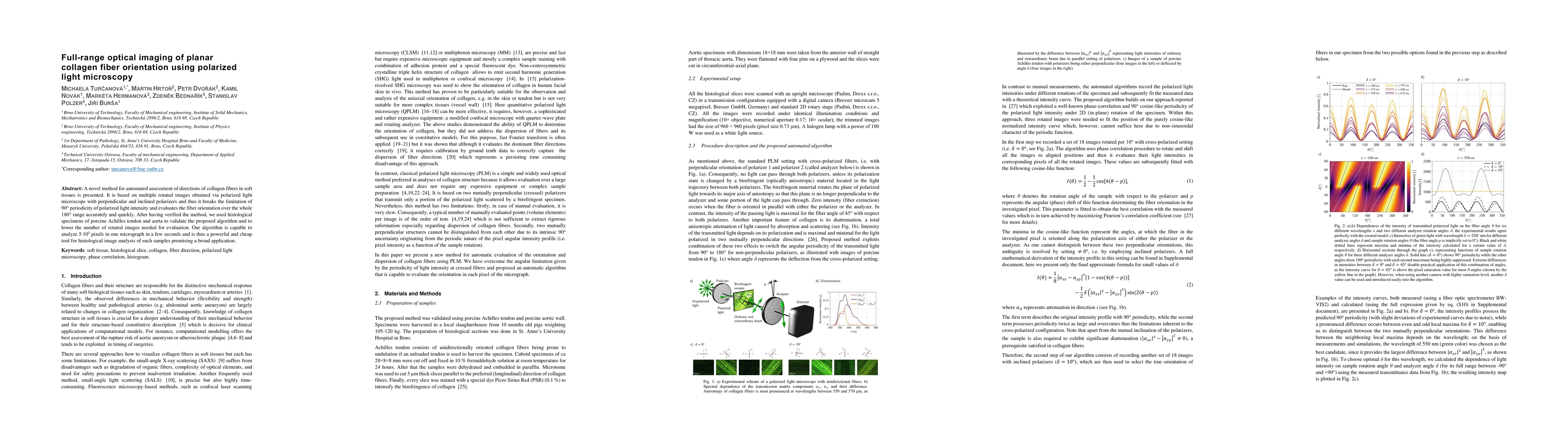
PDF Preview
Key Terms
Citation Network
Current paper (gray), citations (green), references (blue)
Display is limited for performance on very large graphs.
Discussion 0