This study presents a deep learning-based method using U-net to fully automate the analysis of muscle architecture from B-mode ultrasound images, achieving similar accuracy to manual and existing semi-automated methods while significantly reducing analysis time. The method is open source and can estimate fascicle length, pennation angle, and muscle thickness from both single images and videos.
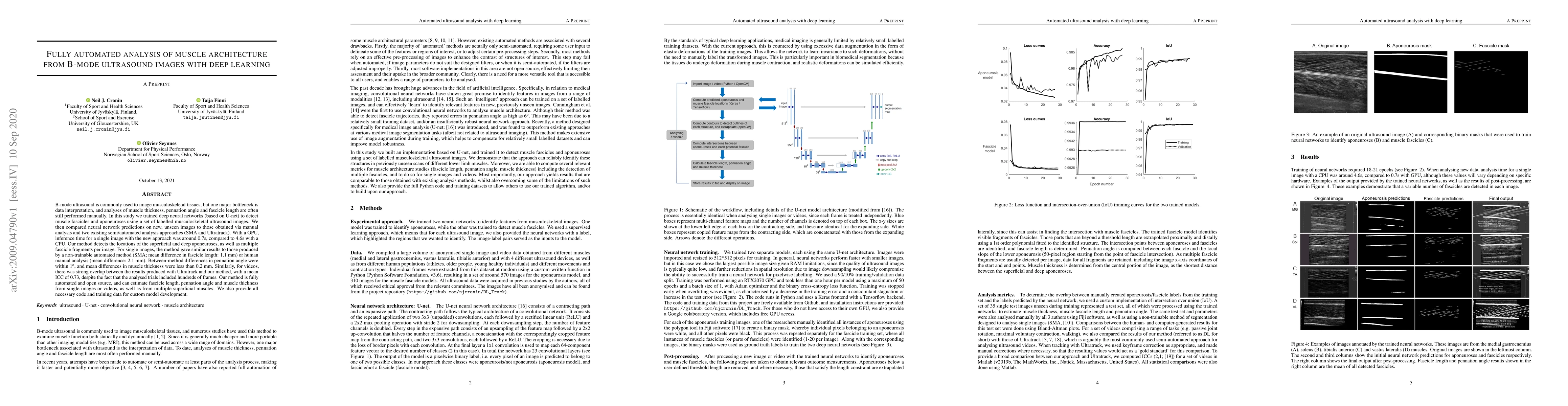
B-mode ultrasound is commonly used to image musculoskeletal tissues, but one major bottleneck is data interpretation, and analyses of muscle thickness, pennation angle and fascicle length are often still performed manually. In this study we trained deep neural networks (based on U-net) to detect muscle fascicles and aponeuroses using a set of labelled musculoskeletal ultrasound images. We then compared neural network predictions on new, unseen images to those obtained via manual analysis and two existing semi/automated analysis approaches (SMA and Ultratrack). With a GPU, inference time for a single image with the new approach was around 0.7s, compared to 4.6s with a CPU. Our method detects the locations of the superficial and deep aponeuroses, as well as multiple fascicle fragments per image. For single images, the method gave similar results to those produced by a non-trainable automated method (SMA; mean difference in fascicle length: 1.1 mm) or human manual analysis (mean difference: 2.1 mm). Between-method differences in pennation angle were within 1$^\circ$, and mean differences in muscle thickness were less than 0.2 mm. Similarly, for videos, there was strong overlap between the results produced with Ultratrack and our method, with a mean ICC of 0.73, despite the fact that the analysed trials included hundreds of frames. Our method is fully automated and open source, and can estimate fascicle length, pennation angle and muscle thickness from single images or videos, as well as from multiple superficial muscles. We also provide all necessary code and training data for custom model development.
This graph shows how this paper is connected to other research. The current paper is shown in dark gray, papers that cite it are in green, and papers it references are in blue. You can:
• Hover over any node to see the paper title
• Click a node to view that paper's details
• Drag nodes to rearrange the graph
• Use the zoom controls (bottom right) to zoom in/out
Title | Authors | Year | Actions |
---|---|---|---|
Quadriceps Muscle Geometry and Strength Throughout Maturation in National-Level Male Soccer Players: A Cross-Sectional Study
|
P. Ritsche, Ralf Roth, Thomas Bernhard, Lukas Nebiker, Eric Lichtenstein, M. Franchi, Jörg Spörri, Oliver Faude
|
2024
|
View Paper |
Muscle Architecture Parameters Inferred from Simulated Single-Element Ultrasound Traces
|
Letizia Gionfrida, Daekyum Kim, Yichu Jin, Conor J. Walsh, Robert D. Howe
|
2024
|
View Paper |
Neuromuscular Modeling for Locomotion with Wearable Assistive Robots - A primer
|
M. Refai, Huawei Wang, A. Gogeascoechea, Rafael Ornelas Kobayashi, L. A. Gaudio, Federica Damonte, G. Durandau, H. V. D. Kooij, U. Yavuz, Massimo Sartori
|
2024
|
View Paper |
RFF-Net: A Refined Feature Feedback Network for Muscle Ultrasound Image Segmentation with Feature Subtraction and Deep Supervision
|
Weida Xie, Ruina Zhao, Tianxiang Li, Deqiang Xiao, Baoting Wang, Hong Song, Meng Yang, Jian Yang
|
2024
|
View Paper |
Improved Fascicle Length Estimates From Ultrasound Using a U-net-LSTM Framework
|
Letizia Gionfrida, R. Nuckols, Conor J. Walsh, Robert D. Howe
|
2023
|
View Paper |
Automated Method for Tracking Human Muscle Architecture on Ultrasound Scans during Dynamic Tasks
|
Saru Meena Ramu, P. Chatzistergos, N. Chockalingam, A. Arampatzis, C. Maganaris
|
2022
|
View Paper |
DeepACSA: Automatic Segmentation of Cross-Sectional Area in Ultrasound Images of Lower Limb Muscles Using Deep Learning
|
P. Ritsche, Philipp Wirth, N. Cronin, F. Sarto, M. Narici, O. Faude, M. Franchi
|
2022
|
View Paper |
Automatic Extraction of Muscle Fascicle Pennation Angle from Raw Ultrasound Data
|
Soley Hafthorsdottir, S. Vostrikov, A. Cossettini, Michael Rieder, Christoph Leitner, Michele Magno, L. Benini
|
2022
|
View Paper |
A Hybrid Method for Ultrasound-Based Tracking of Skeletal Muscle Architecture
|
J. Verheul, S. Yeo
|
2022
|
View Paper |
Fully Automatic Analysis of Muscle B-Mode Ultrasound Images Based on the Deep Residual Shrinkage U-Net
|
Weimin Zheng, Linxueying Zhou, Qing-wei Chai, Jianguo Xu, Shangkun Liu
|
2022
|
View Paper |
DeepACSA: Automatic segmentation of anatomical cross-sectional area in ultrasound images of human lower limb muscles using deep learning
|
P. Ritsche, P. Wirth, N. Cronin, F. Sarto, M. Narici, O. Faude, M. Franchi
|
2021
|
View Paper |
A Human-Centered Machine-Learning Approach for Muscle-Tendon Junction Tracking in Ultrasound Images
|
Christoph Leitner, Robert Jarolim, Bernhard Englmair, A. Kruse, Karen Andrea Lara Hernandez, A. Konrad, Eric Su, Jorg Schrottner, L. Kelly, G. Lichtwark, M. Tilp, Christian F. Baumgartner
|
2021
|
View Paper |
The Influence of Sonographer Experience on Skeletal Muscle Image Acquisition and Analysis
|
J. Carr, Gena R. Gerstner, C. Voskuil, Joel E. Harden, Dustin Dunnick, Kristin Badillo, J. Pagan, K. K. Harmon, R. M. Girts, Jonathan P. Beausejour, M. Stock
|
2021
|
View Paper |
Semi-automated Tracing of Hamstring Muscle Architecture for B-mode Ultrasound Images
|
K. Cronin, E. Delahunt, S. Foley, G. de Vito, Conor McCarthy, S. Cournane
|
2021
|
View Paper |
Deep learning segmentation of transverse musculoskeletal ultrasound images for neuromuscular disease assessment
|
Francesco Marzola, N. Alfen, J. Doorduin, K. Meiburger
|
2021
|
View Paper |
ACSAuto-semi-automatic assessment of human vastus lateralis and rectus femoris cross-sectional area in ultrasound images
|
P. Ritsche, Philipp Wirth, M. Franchi, O. Faude
|
2021
|
View Paper |
A Deep Learning Localization Method for Measuring Abdominal Muscle Dimensions in Ultrasound Images
|
Alzayat Saleh, I. Laradji, C. Lammie, David Vázquez, C. Flavell, M. Azghadi
|
2021
|
View Paper |
Automatic Measurement of Pennation Angle from Ultrasound Images using Resnets
|
Wei-min Zheng, Shangkun Liu, Qing-Wei Chai, Jeng‐Shyang Pan, S. Chu
|
2021
|
View Paper |
Machine learning to extract muscle fascicle length changes from dynamic ultrasound images in real-time
|
Luis G. Rosa, Jonathan S. Zia, O. Inan, G. Sawicki
|
2021
|
View Paper |
A Muscle Pennation Angle Estimation Framework From Raw Ultrasound Data for Wearable Biomedical Instrumentation
|
S. Vostrikov, T. Ingolfsson, Soley Hafthorsdottir, Christoph Leitner, Michele Magno, Luca Benini, Andrea Cossettini
|
2024
|
View Paper |
Published: 2019-06-03
Published: 2020-06-12
Ouwen Huang, Reed Chen, Courtney Trutna Paley, Wren Wightman, Lisa Hobson-Webb, Yohei Harada, Felix Jin, Mark Palmeri, Kathryn Nightingale
Published: 2024-12-07
Comments (0)