The paper presents a fully-automated deep learning-powered system (ECONIB) for DCE-MRI analysis of brain tumors, validated against benchmark (BraTS'17 for tumor segmentation) and clinical (44 low-grade glioma patients) data.
Fully-automated deep learning-powered system for DCE-MRI analysis of brain tumors
Publication
Metrics
AI Quick Summary
A fully-automated deep learning system analyzes brain tumor DCE-MRI scans with high accuracy and speed, reducing manual extraction errors and increasing diagnostic yield.
Quick Answers
What is "Fully-automated deep learning-powered system for DCE-MRI analysis of brain tumors" about?
A fully-automated deep learning system analyzes brain tumor DCE-MRI scans with high accuracy and speed, reducing manual extraction errors and increasing diagnostic yield.
What methodology did the authors use?
The paper presents a fully-automated deep learning-powered system (ECONIB) for DCE-MRI analysis of brain tumors, validated against benchmark (BraTS'17 for tumor segmentation) and clinical (44 low-grade glioma patients) data. More in Methodology →
What are the key results?
ECONIB delivers state-of-the-art results in terms of segmentation accuracy and contrast-concentration fitting. — The system processes an entire input DCE-MRI study in less than 3 minutes using a single GPU. More in Key Results →
Why is this work significant?
ECONIB offers a faster, accurate, and automated solution for DCE-MRI analysis, which is crucial for diagnosis and grading of brain tumors, potentially improving patient care and treatment planning. More in Significance →
What are the main limitations?
The study did not report precision for the validation set. — Performance on smaller and less pronounced lesions might be suboptimal. More in Limitations →
Abstract
Dynamic contrast-enhanced magnetic resonance imaging (DCE-MRI) plays an important role in diagnosis and grading of brain tumor. Although manual DCE biomarker extraction algorithms boost the diagnostic yield of DCE-MRI by providing quantitative information on tumor prognosis and prediction, they are time-consuming and prone to human error. In this paper, we propose a 
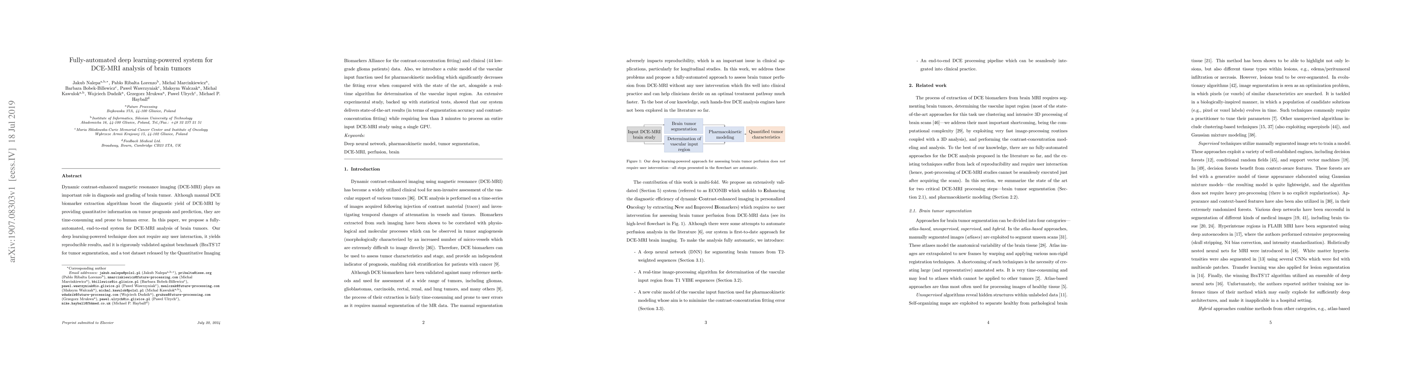
fully-automated, end-to-end system for DCE-MRI analysis of brain tumors. Our deep learning-powered technique does not require any user interaction, it yields reproducible results, and it is rigorously validated against benchmark (BraTS'17 for tumor segmentation, and a test dataset released by the Quantitative Imaging Biomarkers Alliance for the contrast-concentration fitting) and clinical (44 low-grade glioma patients) data. Also, we introduce a cubic model of the vascular input function used for pharmacokinetic modeling which significantly decreases the fitting error when compared with the state of the art, alongside a real-time algorithm for determination of the vascular input region. An extensive experimental study, backed up with statistical tests, showed that our system delivers state-of-the-art results (in terms of segmentation accuracy and contrast-concentration fitting) while requiring less than 3 minutes to process an entire input DCE-MRI study using a single GPU.
Key Findings, in focus
Seven facets of this paper, analysed and brought into focus by AI.
ECONIB offers a faster, accurate, and automated solution for DCE-MRI analysis, which is crucial for diagnosis and grading of brain tumors, potentially improving patient care and treatment planning.
- ECONIB delivers state-of-the-art results in terms of segmentation accuracy and contrast-concentration fitting.
- The system processes an entire input DCE-MRI study in less than 3 minutes using a single GPU.
- A cubic model of the vascular input function (VIF) significantly decreases fitting error compared to state-of-the-art models.
- Real-time algorithm for determination of the vascular input region.
- Robust performance across different MRI data and significantly faster training and deployment compared to top-performing segmentation engines.
ECONIB offers a faster, accurate, and automated solution for DCE-MRI analysis, which is crucial for diagnosis and grading of brain tumors, potentially improving patient care and treatment planning.
ECONIB introduces a novel cubic model for VIF, a real-time algorithm for vascular input region determination, and a deep learning-powered segmentation method that outperforms existing techniques in speed and accuracy.
ECONIB stands out by combining a deep learning-based segmentation approach with a novel cubic VIF model, resulting in a fully automated, accurate, and efficient DCE-MRI analysis system for brain tumors.
- The study did not report precision for the validation set.
- Performance on smaller and less pronounced lesions might be suboptimal.
- Generalization to diverse tumor shapes and sizes needs further validation.
- Validation of DCE-MRI biomarkers extracted by ECONIB for enhanced diagnostic efficiency.
- Comparison of ECONIB biomarkers with those from well-established software (Tissue4D, Siemens) to bring new value to clinical practice.
- Exploration of additional quantitative DCE-MRI features (histogram- or texture-based) for improved analysis.
Impact
Paper Details
PDF Preview
Key Terms
Citation Network
Current paper (gray), citations (green), references (blue)
Display is limited for performance on very large graphs.
Discussion 0