Fully Automated Tree Topology Estimation and Artery-Vein Classification
Publication
Metrics
AI Quick Summary
This paper introduces a fully automated, graph-based method for extracting retinal vascular topology from a single color fundus image, using high-level operations to correct errors and achieve state-of-the-art results in retinal artery-vein classification across multiple datasets. The method combines vessel segmentation and a novel cost function to optimize the vascular graph, validated through ablation studies.
Paper Preview
Abstract
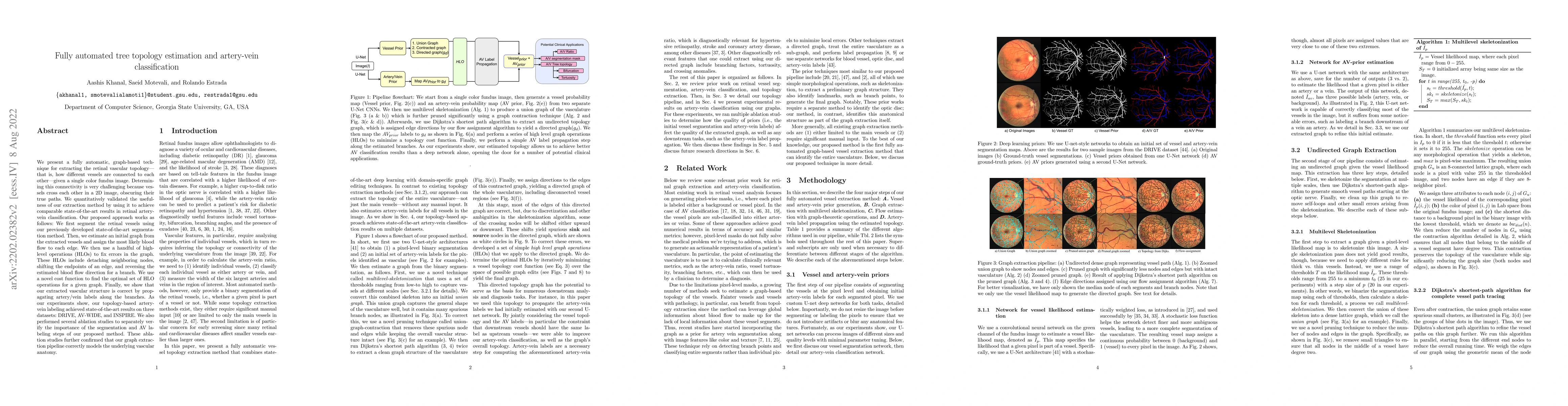
We present a fully automatic, graph-based technique for extracting the retinal vascular topology -- that is, how different vessels are connected to each other -- given a single color fundus image. Determining this connectivity is very challenging because vessels cross each other in a 2D image, obscuring their true paths. We quantitatively validated the usefulness of our extraction method by using it to achieve comparable state-of-the-art results in retinal artery-vein classification. Our proposed approach works as follows: We first segment the retinal vessels using our previously developed state-of-the-art segmentation method. Then, we estimate an initial graph from the extracted vessels and assign the most likely blood flow to each edge. We then use a handful of high-level operations (HLOs) to fix errors in the graph. These HLOs include detaching neighboring nodes, shifting the endpoints of an edge, and reversing the estimated blood flow direction for a branch. We use a novel cost function to find the optimal set of HLO operations for a given graph. Finally, we show that our extracted vascular structure is correct by propagating artery/vein labels along the branches. As our experiments show, our topology-based artery-vein labeling achieved state-of-the-art results on three datasets: DRIVE, AV-WIDE, and INSPIRE. We also performed several ablation studies to separately verify the importance of the segmentation and AV labeling steps of our proposed method. These ablation studies further confirmed that our graph extraction pipeline correctly models the underlying vascular anatomy.
AI Key Findings
Get AI-generated insights about this paper's methodology, results, significance, and more — seven facets brought into focus.
Impact
Paper Details
Authors
PDF Preview
Key Terms
Citation Network
Current paper (gray), citations (green), references (blue)
Display is limited for performance on very large graphs.
Discussion 0