This paper presents a fully automatic segmentation pipeline for myocardial contrast echocardiography using random forests guided by a statistical shape model, which improves accuracy by incorporating shape priors and refining segmentations, outperforming other methods in clinical data evaluation. The approach also includes a bounding box detection and extends to 2D+t sequences for temporal consistency.
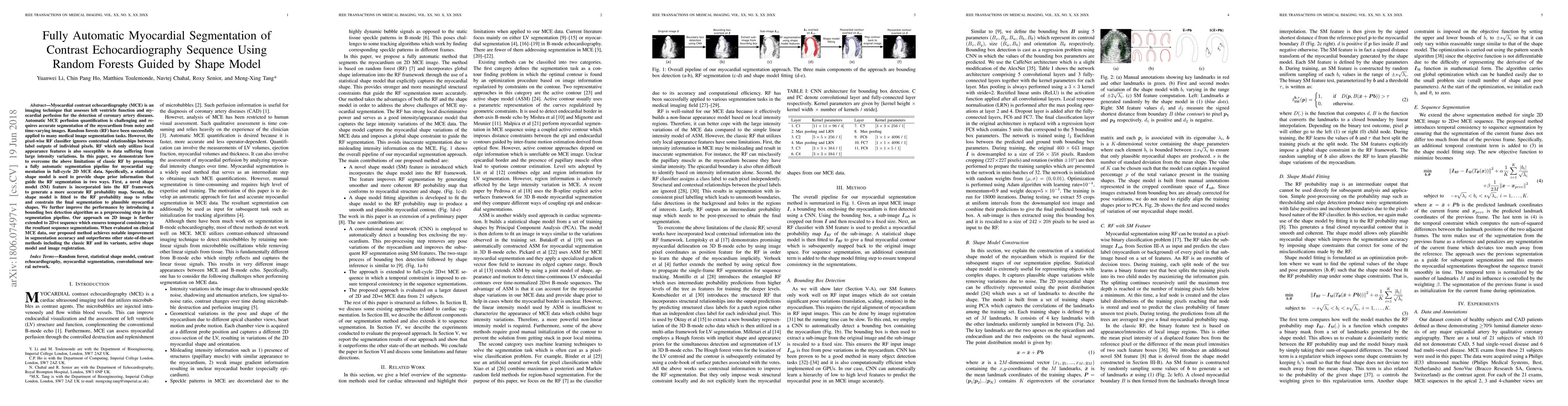
Myocardial contrast echocardiography (MCE) is an imaging technique that assesses left ventricle function and myocardial perfusion for the detection of coronary artery diseases. Automatic MCE perfusion quantification is challenging and requires accurate segmentation of the myocardium from noisy and time-varying images. Random forests (RF) have been successfully applied to many medical image segmentation tasks. However, the pixel-wise RF classifier ignores contextual relationships between label outputs of individual pixels. RF which only utilizes local appearance features is also susceptible to data suffering from large intensity variations. In this paper, we demonstrate how to overcome the above limitations of classic RF by presenting a fully automatic segmentation pipeline for myocardial segmentation in full-cycle 2D MCE data. Specifically, a statistical shape model is used to provide shape prior information that guide the RF segmentation in two ways. First, a novel shape model (SM) feature is incorporated into the RF framework to generate a more accurate RF probability map. Second, the shape model is fitted to the RF probability map to refine and constrain the final segmentation to plausible myocardial shapes. We further improve the performance by introducing a bounding box detection algorithm as a preprocessing step in the segmentation pipeline. Our approach on 2D image is further extended to 2D+t sequence which ensures temporal consistency in the resultant sequence segmentations. When evaluated on clinical MCE data, our proposed method achieves notable improvement in segmentation accuracy and outperforms other state-of-the-art methods including the classic RF and its variants, active shape model and image registration.
Get AI-generated insights about this paper's methodology, results, and significance.
Current paper (gray), citations (green), references (blue)
Lei Li, Xiahai Zhuang, Shan Yang et al.
Title | Authors | Year | Actions |
---|---|---|---|
CMR-BENet: A confidence map refinement boundary enhancement network for left ventricular myocardium segmentation.
|
Qi Yu, Hongxia Ning, Jinzhu Yang, Chen Li, Yiqiu Qi, Ming Qu, Honghe Li, Song Sun, Peng Cao, Chaolu Feng
|
2024
|
View Paper |
TreeFormer: Single-view Plant Skeleton Estimation via Tree-constrained Graph Generation
|
Xinpeng Liu, Hiroaki Santo, Yosuke Toda, Fumio Okura
|
2024
|
View Paper |
SPGNet: A Shape-prior Guided Network for Medical Image Segmentation
|
Zhengxuan Song, Xun Liu, Wenhao Zhang, Yongyi Gong, T. Hao, Kun Zeng
|
2024
|
View Paper |
Dual-Branch TransV-Net for 3-D Echocardiography Segmentation
|
Jiapeng Zhang, Yongxiong Wang, Lijun Chen, Jinlong Liu, Sunjie Zhang, Zhiqun Pan, Zhe Wang, Zhenhui Tang, Ying Guo
|
2023
|
View Paper |
Simultaneous Hip Implant Segmentation and Gruen Landmarks Detection
|
Asma Alzaid, B. Lineham, S. Dogramadzi, H. Pandit, A. Frangi, S. Xie
|
2023
|
View Paper |
Toward automated left ventricle segmentation of whole-cardiac-cycle MR images via Contextual-Feature-Induced Semantic Flow Propagation Network
|
Menke Li, Jinping Liu, Subo Gong, Hadi Jahanshahi
|
2023
|
View Paper |
Topology-sensitive weighting model for myocardial segmentation
|
Song Sun, Yonghuai Wang, Jinzhu Yang, Yong Feng, Lingzhi Tang, Shuo Liu, Hongxia Ning
|
2023
|
View Paper |
DSANet: Dual-Branch Shape-Aware Network for Echocardiography Segmentation in Apical Views
|
Guangquan Zhou, Wen-Bo Zhang, Zhongqing Shi, Zhanru Qi, Kaini Wang, Hong Song, Jing Yao, Yang Chen
|
2023
|
View Paper |
A Review of Medical Diagnostic Video Analysis Using Deep Learning Techniques
|
Moomal Farhad, M. Masud, A. Beg, Amir Ahmad, L. Ahmed
|
2023
|
View Paper |
Semantic segmentation method for myocardial contrast echocardiogram based on DeepLabV3+ deep learning architecture.
|
Huan Cheng, Jucheng Zhang, Yinglan Gong, Z. Pu, Jun Jiang, Yonghua Chu, Ling Xia
|
2023
|
View Paper |
A Study on Various Segmentation Approaches using Echocardiogram images for promoting Heart Disease Detection
|
Professor, Nagashettappa Biradar, Bheemanna Khandre, A. Jadhav, Hamjad Ali Umachagi
|
2022
|
View Paper |
Improved Segmentation of Echocardiography With Orientation-Congruency of Optical Flow and Motion-Enhanced Segmentation
|
Wufeng Xue, Heng Cao, Junqiang Ma, T. Bai, Tianfu Wang, Dong Ni
|
2022
|
View Paper |
Semi-automatic Segmentation of the Myocardium in High-Frame Rate and Clinical Contrast Echocardiography Images
|
S. Sze, Oscar Bates, M. Toulemonde, M. Tang, Gabriel Bioh, R. Senior
|
2022
|
View Paper |
Assessment and Treatment for Coronary Microvascular Dysfunction by Contrast Enhanced Ultrasound
|
Junzhen Zhan, Longhe Zhong, Juefei Wu
|
2022
|
View Paper |
Optimization algorithm of CT image edge segmentation using improved convolution neural network
|
Xiaojuan Wang, Yuntao Wei
|
2022
|
View Paper |
Deep learning applications in myocardial perfusion imaging, a systematic review and meta-analysis
|
E. Alskaf, Utkarsh Dutta, C. Scannell, A. Chiribiri
|
2022
|
View Paper |
Invited:Hardware-aware Real-time Myocardial Segmentation Quality Control in Contrast Echocardiography
|
Dewen Zeng, Yukun Ding, Haiyun Yuan, Meiping Huang, Xiaowei Xu, Zhuang Jian, Jingtong Hu, Yiyu Shi
|
2021
|
View Paper |
Contrastive Learning for View Classification of Echocardiograms
|
A. Chartsias, Shan Gao, A. Mumith, Jorge Oliveira, K. Bhatia, Bernhard Kainz, A. Beqiri
|
2021
|
View Paper |
Echocardiogram segmentation using active shape model and mean squared eigenvalue error
|
Yasser Ali, S. Beheshti, F. Janabi-Sharifi
|
2021
|
View Paper |
CAD based product packaging design scheme based on deformed shape image detection and quality assessment
|
Xiaofeng Wang
|
2021
|
View Paper |
Invasive and Non-Invasive Imaging for Ischaemia with No Obstructive Coronary Artery Disease
|
M. Ng, Hok Shing Tang, L. Fong, Victor Chan, R. Senior, D. Pennell
|
2021
|
View Paper |
Amniotic fluid segmentation based on pixel classification using local window information and distance angle pixel
|
Desiana Wulaning Ayu, S. Hartati, Aina Musdholifah, D. Nurdiati
|
2021
|
View Paper |
A deep learning approach with temporal consistency for automatic myocardial segmentation of quantitative myocardial contrast echocardiography
|
Mingqi Li, Dewen Zeng, Qiu Xie, Ruixue Xu, Yu Wang, Dunliang Ma, Yiyu Shi, Xiaowei Xu, Meiping Huang, Hongwen Fei
|
2021
|
View Paper |
A Survey on Shape-Constraint Deep Learning for Medical Image Segmentation
|
Simon Bohlender, Ilkay Öksüz, A. Mukhopadhyay
|
2021
|
View Paper |
Stress CMR in Known or Suspected CAD: Diagnostic and Prognostic Role
|
F. Baessato, M. Guglielmo, G. Muscogiuri, A. Baggiano, L. Fusini, Stefano Scafuri, M. Babbaro, R. Mollace, A. Collevecchio, A. Guaricci, G. Pontone
|
2021
|
View Paper |
Deep learning in spatiotemporal cardiac imaging: A review of methodologies and clinical usability
|
Karen Andrea Lara Hernandez, T. Rienmüller, D. Baumgartner, C. Baumgartner
|
2020
|
View Paper |
Shape prior model via dual subspace segment projection learning
|
G. Belous, Andrew Busch, Yongsheng Gao
|
2020
|
View Paper |
Deep RetinaNet for Dynamic Left Ventricle Detection in Multiview Echocardiography Classification
|
Meijun Yang, Xiaoyan Xiao, Zhi Liu, Longkun Sun, W. Guo, Li-zhen Cui, Dianmin Sun, Pengfei Zhang, Guang Yang
|
2020
|
View Paper |
Vessel Segmentation of X-Ray Coronary Angiographic Image Sequence
|
Shaoyan Xia, Haogang Zhu, Xiaoli Liu, Ming Gong, Xiaoyong Huang, Lei Xu, Hongjia Zhang, Jialong Guo
|
2020
|
View Paper |
Object Segmentation with Deep Neural Nets Coupled with a Shape Prior, When Learning From a Training set of Limited Quality and Small Size
|
Saurabh J. Shigwan, Akshay V. Gaikwad, Suyash P. Awate
|
2020
|
View Paper |
PRF-RW: a progressive random forest-based random walk approach for interactive semi-automated pulmonary lobes segmentation
|
Qiang Li, Lei Chen, Xiangju Li, Xiaofeng Lv, Shuyue Xia, Yan Kang
|
2020
|
View Paper |
Clinical quantitative cardiac imaging for the assessment of myocardial ischaemia
|
M. Dewey, M. Siebes, M. Kachelriess, K. Kofoed, P. Maurovich-Horvát, K. Nikolaou, Wenjia Bai, A. Kofler, R. Manka, S. Kozerke, A. Chiribiri, T. Schaeffter, Florian Michallek, F. Bengel, S. Nekolla, P. Knaapen, M. Lubberink, R. Senior, M. Tang, J. Piek, T. P. van de Hoef, Johannes Martens, L. Schreiber
|
2020
|
View Paper |
Discriminant analysis of mine quake type and intensity based on a deep neural network
|
Hui Liu, Xiaojun Zhang
|
2020
|
View Paper |
Weakly Supervised Deformation Network for 3D Echocardiography Segmentation on Left Ventricle
|
Suyu Dong, Gongning Luo, Naren Wulan, Shaodong Cao, Kuanquan Wang, Henggui Zhang
|
2019
|
View Paper |
Amniotic Fluid Segmentation by Pixel Classification in B-Mode Ultrasound Image for Computer Assisted Diagnosis
|
Desiana Wulaning Ayu, S. Hartati, Aina Musdholifah
|
2019
|
View Paper |
Machine Learning for Data-Driven Discovery: The Rise and Relevance.
|
P. Sengupta, S. Shrestha
|
2019
|
View Paper |
Differentiation of patients with two-dimensional echocardiography false positive non-compaction of ventricular myocardium by contrast echocardiography.
|
Mingjun Tian, Minjuan Zheng, Hui Ma, Yicui Niu, Ying Zhang, Pan Xu
|
2019
|
View Paper |
High-Frame-Rate Contrast Echocardiography Using Diverging Waves: Initial In Vitro and In Vivo Evaluation
|
M. Toulemonde, Yuanwei Li, Shengtao Lin, F. Cordonnier, M. Butler, W. C. Duncan, R. Eckersley, V. Sboros, M. Tang
|
2018
|
View Paper |
ETAB: A Benchmark Suite for Visual Representation Learning in Echocardiography
|
Ahmed M. Alaa, Anthony Philippakis, David A. Sontag
|
2022
|
View Paper |
Amniotic Fluids Classification Using Combination of Rules-Based and Random Forest Algorithm
|
Putu Desiana Wulaning Ayu, S. Hartati, Aina Musdholifah, D. Nurdiati
|
2021
|
View Paper |
Cxnet-M3: A Deep Quintuplet Network for Multi-Lesion Classification in Chest X-Ray Images Via Multi-Label Supervision
|
Shuaijing Xu, Xiaoyilei Yang, Junqi Guo, Hao Wu, Guangzhi Zhang, R. Bie
|
2020
|
View Paper |
CXNet-m1: Anomaly Detection on Chest X-Rays With Image-Based Deep Learning
|
Shuaijing Xu, Hao Wu, R. Bie
|
2019
|
View Paper |
High-FrameRate Contrast Echocardiography using diverging waves : initial in-vitro and in-vivo evaluation
|
M. Toulemonde, S. Lin, F. Cordonnier, M. Butler, C. Duncan, R. Eckersley, V. Sboros, M. Tang
|
2018
|
View Paper |
Coronary heart disease monitoring system based on wireless sensors
|
M. Dweck, S. Mirsadraee, A. Fletcher, C. Lucatelli, D. Patel, E. Beek, D. Newby
|
2018
|
View Paper |
Comments (0)