Fully Automatic Segmentation of Lumbar Vertebrae from CT Images using Cascaded 3D Fully Convolutional Networks
Publication
Metrics
AI Quick Summary
This paper introduces a method for segmenting lumbar vertebrae from CT images using cascaded 3D Fully Convolutional Networks (FCNs). The approach employs a "LocalizationNet" to identify the lumbar region and a "SegmentationNet" for precise pixel-wise segmentation, achieving an average Dice coefficient of 95.77% and a symmetric surface distance of 0.37 mm.
Paper Preview
Abstract
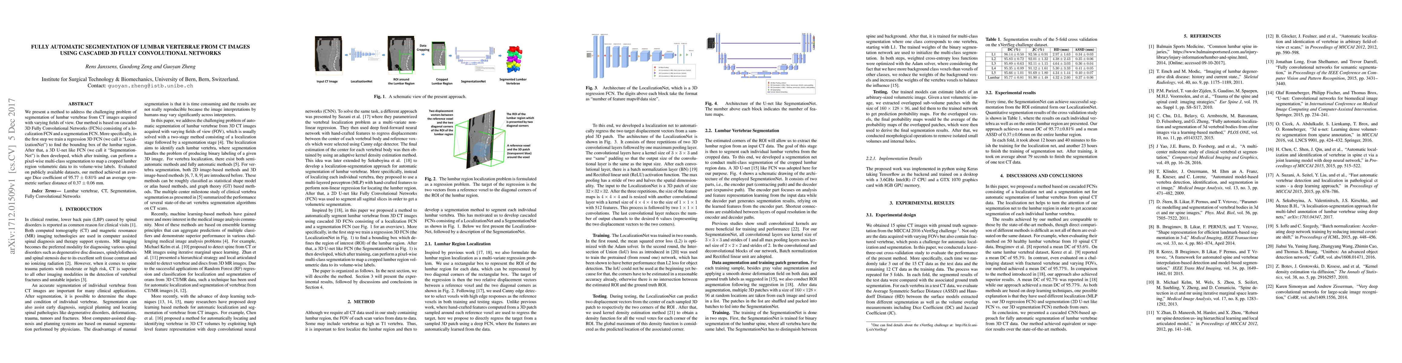
We present a method to address the challenging problem of segmentation of lumbar vertebrae from CT images acquired with varying fields of view. Our method is based on cascaded 3D Fully Convolutional Networks (FCNs) consisting of a localization FCN and a segmentation FCN. More specifically, in the first step we train a regression 3D FCN (we call it "LocalizationNet") to find the bounding box of the lumbar region. After that, a 3D U-net like FCN (we call it "SegmentationNet") is then developed, which after training, can perform a pixel-wise multi-class segmentation to map a cropped lumber region volumetric data to its volume-wise labels. Evaluated on publicly available datasets, our method achieved an average Dice coefficient of 95.77 $\pm$ 0.81% and an average symmetric surface distance of 0.37 $\pm$ 0.06 mm.
AI Key Findings
Get AI-generated insights about this paper's methodology, results, significance, and more — seven facets brought into focus.
Impact
Paper Details
PDF Preview
Key Terms
Citation Network
Current paper (gray), citations (green), references (blue)
Display is limited for performance on very large graphs.
Discussion 0