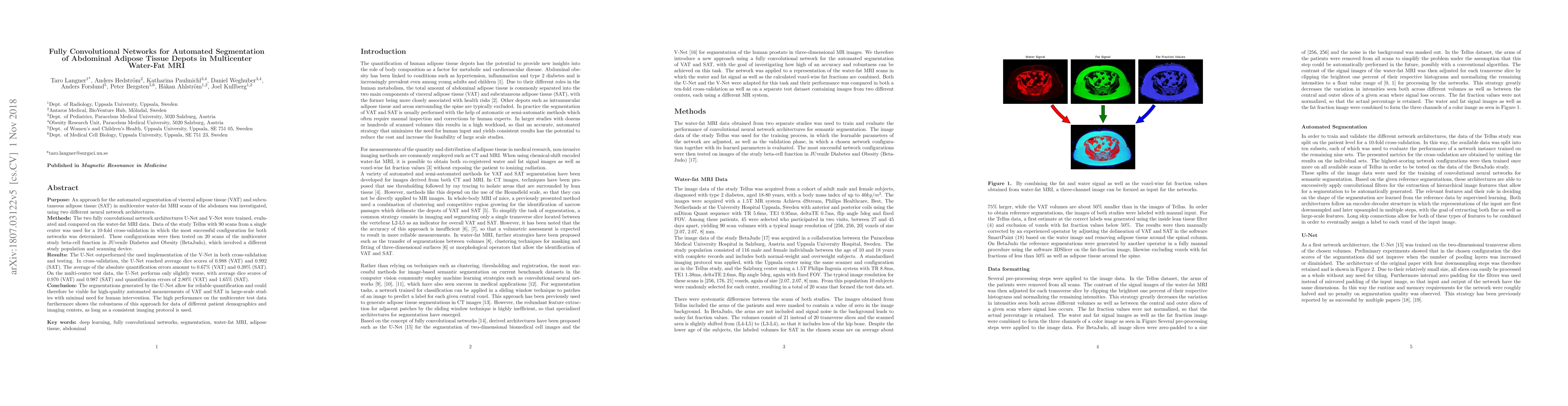
Summary
Purpose: An approach for the automated segmentation of visceral adipose tissue (VAT) and subcutaneous adipose tissue (SAT) in multicenter water-fat MRI scans of the abdomen was investigated, using two different neural network architectures. Methods: The two fully convolutional network architectures U-Net and V-Net were trained, evaluated and compared on the water-fat MRI data. Data of the study Tellus with 90 scans from a single center was used for a 10-fold cross-validation in which the most successful configuration for both networks was determined. These configurations were then tested on 20 scans of the multicenter study beta-cell function in JUvenile Diabetes and Obesity (BetaJudo), which involved a different study population and scanning device. Results: The U-Net outperformed the used implementation of the V-Net in both cross-validation and testing. In cross-validation, the U-Net reached average dice scores of 0.988 (VAT) and 0.992 (SAT). The average of the absolute quantification errors amount to 0.67% (VAT) and 0.39% (SAT). On the multi-center test data, the U-Net performs only slightly worse, with average dice scores of 0.970 (VAT) and 0.987 (SAT) and quantification errors of 2.80% (VAT) and 1.65% (SAT). Conclusion: The segmentations generated by the U-Net allow for reliable quantification and could therefore be viable for high-quality automated measurements of VAT and SAT in large-scale studies with minimal need for human intervention. The high performance on the multicenter test data furthermore shows the robustness of this approach for data of different patient demographics and imaging centers, as long as a consistent imaging protocol is used.
AI Key Findings
Get AI-generated insights about this paper's methodology, results, and significance.
Paper Details
PDF Preview
Key Terms
Citation Network
Current paper (gray), citations (green), references (blue)
Display is limited for performance on very large graphs.
Similar Papers
Found 4 papersAutomatic quantification of abdominal subcutaneous and visceral adipose tissue in children, through MRI study, using total intensity maps and Convolutional Neural Networks
Po-Wah So, José Gerardo Suárez-García, Javier Miguel Hernández-López et al.
| Title | Authors | Year | Actions |
|---|
Comments (0)