Functional Connectivity Dynamics show Resting-State Instability and Rightward Parietal Dysfunction in ADHD

Source: ArXiv

AI Quick Summary

This study reveals that children with ADHD exhibit significantly higher temporal variability in functional connectivity compared to typically developing children, particularly in the cingulo-temporal, cingulo-parietal, fronto-temporal, and fronto-parietal networks, suggesting resting-state instability and rightward parietal dysfunction associated with ADHD.

Share: Share on X LinkedIn

Summary

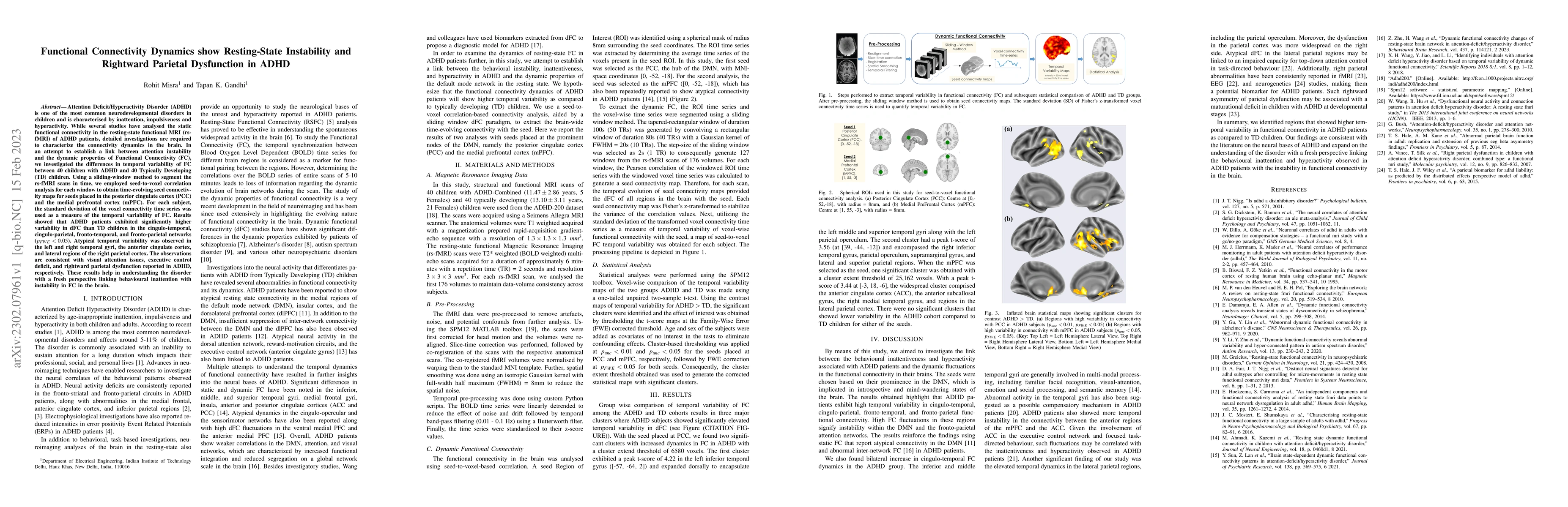
Attention Deficit/Hyperactivity Disorder (ADHD) is one of the most common neurodevelopmental disorders in children and is characterised by inattention, impulsiveness and hyperactivity. While several studies have analysed the static functional connectivity in the resting-state functional MRI (rs-fMRI) of ADHD patients, detailed investigations are required to characterize the connectivity dynamics in the brain. In an attempt to establish a link between attention instability and the dynamic properties of Functional Connectivity (FC), we investigated the differences in temporal variability of FC between 40 children with ADHD and 40 Typically Developing (TD) children. Using a sliding-window method to segment the rs-fMRI scans in time, we employed seed-to-voxel correlation analysis for each window to obtain time-evolving seed connectivity maps for seeds placed in the posterior cingulate cortex (PCC) and the medial prefrontal cortex (mPFC). For each subject, the standard deviation of the voxel connectivity time series was used as a measure of the temporal variability of FC. Results showed that ADHD patients exhibited significantly higher variability in dFC than TD children in the cingulo-temporal, cingulo-parietal, fronto-temporal, and fronto-parietal networks ($p_{FWE} < 0.05$). Atypical temporal variability was observed in the left and right temporal gyri, the anterior cingulate cortex, and lateral regions of the right parietal cortex. The observations are consistent with visual attention issues, executive control deficit, and rightward parietal dysfunction reported in ADHD, respectively. These results help in understanding the disorder with a fresh perspective linking behavioural inattention with instability in FC in the brain.

AI Key Findings

Generated Sep 05, 2025

Methodology

This study used functional magnetic resonance imaging (fMRI) to investigate the neural correlates of attention-deficit/hyperactivity disorder (ADHD). The analysis focused on resting-state functional connectivity in the brain network.

Key Results

  • Temporally unstable functional connectivity was found between the anterior regions of the prefrontal cortex and the anterior cingulate cortex in ADHD individuals
  • Increased temporal dynamics were observed in the lateral parietal regions, which may be related to impaired top-down attention control
  • Rightward asymmetry of parietal dysfunction was consistently reported across studies, suggesting a potential biomarker for ADHD

Significance

This study provides new insights into the neural basis of ADHD and highlights the importance of temporal variability in functional connectivity. The findings have implications for the diagnosis and treatment of ADHD.

Technical Contribution

This study developed a novel approach to analyzing resting-state fMRI data, which can be applied to other neurological disorders

Novelty

The study identified distinct neural signatures for ADHD subtypes, providing new insights into the heterogeneity of the disorder

Limitations

  • The sample size was relatively small, which may limit the generalizability of the results
  • The study relied on resting-state fMRI data, which may not capture task-dependent neural activity

Future Work

  • Investigating the neural correlates of ADHD in larger samples and using more advanced neuroimaging techniques
  • Examining the relationship between temporal variability in functional connectivity and cognitive symptoms in ADHD

Paper Details

PDF Preview

Download PDF

Key Terms

children (0.296)
connectivity (0.283)
temporal (0.243)
variability (0.235)
rs (0.216)
brain (0.189)
patients (0.184)
window (0.183)

Citation Network

Current paper (gray), citations (green), references (blue)

Display is limited for performance on very large graphs.

Current Paper
Citations
References
Click to view
Title Authors Year Actions

Comments (0)

This website uses cookies to ensure you get the best experience. Learn more