Publication
Metrics
AI Quick Summary
This study demonstrates the first successful application of functional magnetic resonance spectroscopy (fMRS) in mice, showing its feasibility in quantifying metabolic variations during visual stimulation. The results reveal significant modulations in metabolites like glutamate, NAAG, PCr, and Cr, highlighting the potential of fMRS for future research in murine models.
Paper Preview
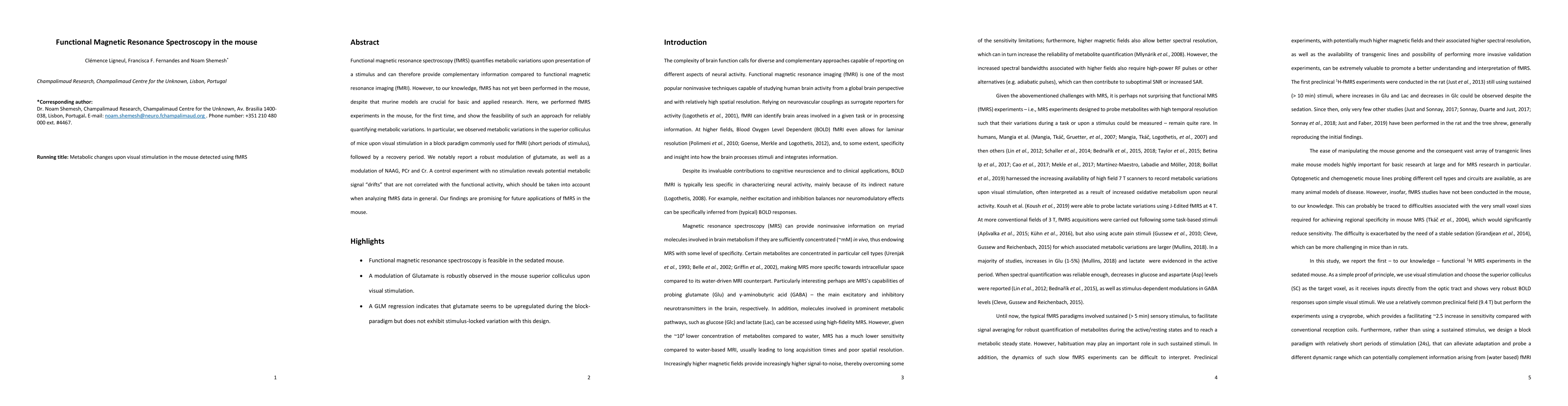
Abstract
Functional magnetic resonance spectroscopy (fMRS) quantifies metabolic variations upon presentation of a stimulus and can therefore provide complementary information compared to functional magnetic resonance imaging (fMRI). However, to our knowledge, fMRS has not yet been performed in the mouse, despite that murine models are crucial for basic and applied research. Here, we performed fMRS experiments in the mouse, for the first time, and show the feasibility of such an approach for reliably quantifying metabolic variations. In particular, we observed metabolic variations in the superior colliculus of mice upon visual stimulation in a block paradigm commonly used for fMRI (short periods of stimulus), followed by a recovery period. We notably report a robust modulation of glutamate, as well as a modulation of NAAG, PCr and Cr. A control experiment with no stimulation reveals potential metabolic signal "drifts" that are not correlated with the functional activity, which should be taken into account when analyzing fMRS data in general. Our findings are promising for future applications of fMRS in the mouse.
AI Key Findings
Get AI-generated insights about this paper's methodology, results, significance, and more — seven facets brought into focus.
Impact
Paper Details
Authors
PDF Preview
Key Terms
Citation Network
Current paper (gray), citations (green), references (blue)
Display is limited for performance on very large graphs.
Discussion 0