Publication
Metrics
AI Quick Summary
It aims to bridge the gap between fundamental concepts in physics and real-world medical uses, making it an accessible resource for STEM students.
Paper Preview
Abstract
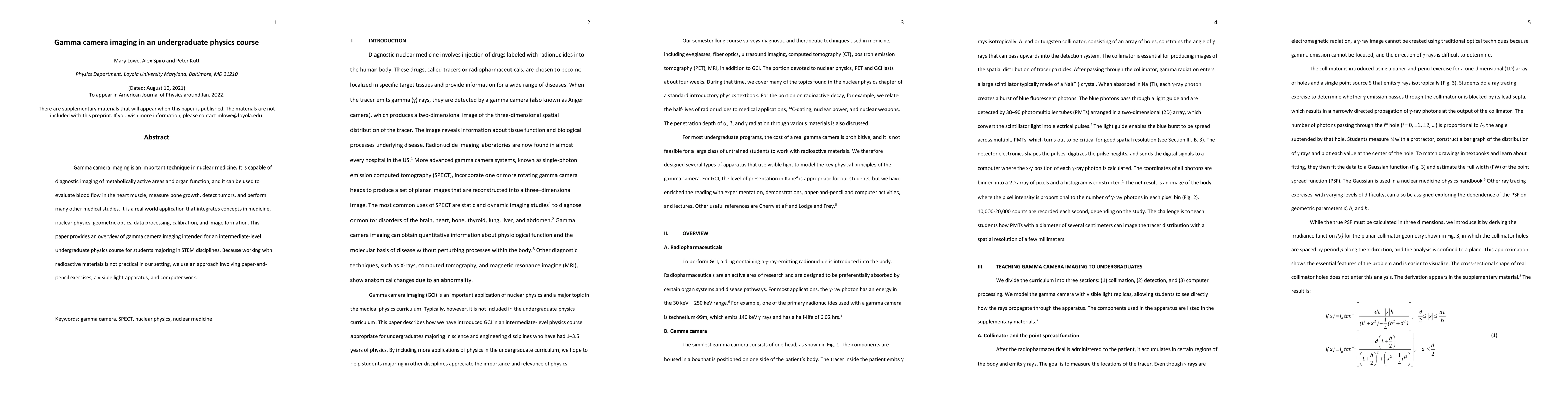
Gamma camera imaging is an important technique in nuclear medicine. It is capable of diagnostic imaging of metabolically active areas and organ function, and it can be used to evaluate blood flow in the heart muscle, measure bone growth, detect tumors, and perform many other medical studies. It is a real world application that integrates concepts in medicine, nuclear physics, geometric optics, data processing, calibration, and image formation. This paper provides an overview of gamma camera imaging intended for an intermediate-level undergraduate physics course for students majoring in STEM disciplines. Because working with radioactive materials is not practical in our setting, we use an approach involving paper-and-pencil exercises, visible light apparatus, and computer work.
AI Key Findings
Get AI-generated insights about this paper's methodology, results, significance, and more — seven facets brought into focus.
Impact
Paper Details
Authors
PDF Preview
Key Terms
Citation Network
Current paper (gray), citations (green), references (blue)
Display is limited for performance on very large graphs.
Discussion 0