Generative Model-Based Ischemic Stroke Lesion Segmentation

PDF

Publication

Published: Jun 07, 2019
Categories: eess.IV, cs.CV, cs.LG

Metrics

Source: ArXiv

Quick Actions

View PDF

AI Quick Summary

This paper proposes a generative model-based framework for ischemic stroke lesion segmentation using CT perfusion data, which includes an extractor, a generator, and a segmentor. The method won first place in the 2018 Ischemic Stroke Lesion Segmentation Challenge, demonstrating effective segmentation using a novel pixel-region loss function.

Quick Answers

What methodology did the authors use?

A novel generative model-based end-to-end segmentation framework is proposed for ischemic stroke lesion segmentation using CT perfusion images. More in Methodology →

What are the key results?

Achieved a Dice coefficient of 62.40% in cross-validation stage — Won the first place in the test stage of 2018 ischemic stroke lesions segmentation challenge More in Key Results →

Why is this work significant?

This research is important because it addresses the highly unbalanced problem of accurate stroke lesion segmentation using CT perfusion images. More in Significance →

What are the main limitations?

The proposed framework may not generalize well to unseen data or patients — The use of deep learning-based methods may require significant computational resources More in Limitations →

Paper Preview

Abstract

CT perfusion (CTP) has been used to triage ischemic stroke patients in the early stage, because of its speed, availability, and lack of contraindications. Perfusion parameters including cerebral blood volume (CBV), cerebral blood flow (CBF), mean transit time (MTT) and time of peak (Tmax) could also be computed from CTP data. However, CTP data or the perfusion parameters, are ambiguous to locate the infarct core or tissue at risk (penumbra), which is normally confirmed by the follow-up Diffusion Weighted Imaging (DWI) or perfusion 
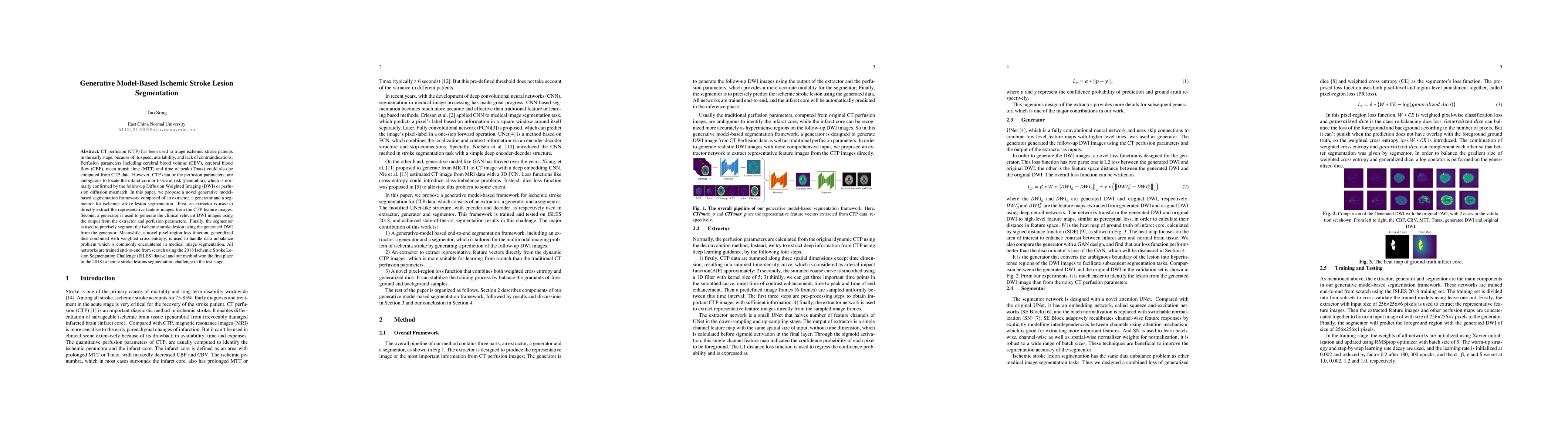
diffusion mismatch. In this paper, we propose a novel generative modelbased segmentation framework composed of an extractor, a generator and a segmentor for ischemic stroke lesion segmentation. First, an extractor is used to directly extract the representative feature images from the CTP feature images. Second, a generator is used to generate the clinical relevant DWI images using the output from the extractor and perfusion parameters. Finally, the segmentor is used to precisely segment the ischemic stroke lesion using the generated DWI from the generator. Meanwhile, a novel pixel-region loss function, generalized dice combined with weighted cross entropy, is used to handle data unbalance problem which is commonly encountered in medical image segmentation. All networks are trained end-to-end from scratch using the 2018 Ischemic Stroke Lesion Segmentation Challenge (ISLES) dataset and our method won the first place in the 2018 ischemic stroke lesions segmentation challenge in the test stage.

AI Key Findings

Generated Sep 04, 2025

Methodology — What approach did the authors take?

A novel generative model-based end-to-end segmentation framework is proposed for ischemic stroke lesion segmentation using CT perfusion images.

Key Results — What are the main findings?

  • Achieved a Dice coefficient of 62.40% in cross-validation stage
  • Won the first place in the test stage of 2018 ischemic stroke lesions segmentation challenge
  • Improved upon existing methods with robust performance on challenging data

Significance — Why does this research matter?

This research is important because it addresses the highly unbalanced problem of accurate stroke lesion segmentation using CT perfusion images.

Technical Contribution — What is the technical contribution?

A novel generative model-based approach is proposed, which combines an extractor, a generator, and a segmentor to achieve accurate segmentation.

Novelty — What is new about this work?

This work is novel because it proposes a multi-stage framework that leverages deep learning-based methods to generate clinically relevant images for segmentation.

Limitations — What are the limitations of this study?

  • The proposed framework may not generalize well to unseen data or patients
  • The use of deep learning-based methods may require significant computational resources

Future Work — What did the authors propose for future work?

  • Exploring the usage of this framework in clinical workflow of stroke patients
  • Replacing devolution-based perfusion parameters with deep learning-based perfusion feature images

Paper Details

Paper ID: 1906.02392
License: http://arxiv.org/licenses/nonexclusive-distrib/1.0/
Categories:
eess.IV cs.CV cs.LG

How to Cite This Paper

BibTeX
@article{anon2019generative,
  title         = {Generative Model-Based Ischemic Stroke Lesion Segmentation},
  year          = {2019},
  eprint        = {1906.02392},
  archivePrefix = {arXiv},
  primaryClass  = {eess.IV},
}
APA 7
(2019). Generative Model-Based Ischemic Stroke Lesion Segmentation. arXiv. https://arxiv.org/abs/1906.02392
MLA 9
"Generative Model-Based Ischemic Stroke Lesion Segmentation." arXiv, 2019, arxiv.org/abs/1906.02392.

Key Terms

segmentation (0.282)
generator (0.240)
stage (0.179)
images (0.177)
weighted (0.170)
diffusion (0.163)
challenge (0.158)
used (0.150)

Citation Network

Current paper (gray), citations (green), references (blue)

Display is limited for performance on very large graphs.

Current Paper
Citations
References
Click to view
Title Authors Year Actions

Comments (0)

This website uses cookies to ensure you get the best experience. Learn more