Generative Synthetic Augmentation using Label-to-Image Translation for Nuclei Image Segmentation
Publication
Metrics
AI Quick Summary
This paper proposes a generative synthetic augmentation method using label-to-image translation to enhance nuclei image segmentation in digital pathology. The approach aims to improve segmentation accuracy by augmenting rare tumor images with synthetic data, demonstrating improved performance over traditional segmentation algorithms.
Paper Preview
Abstract
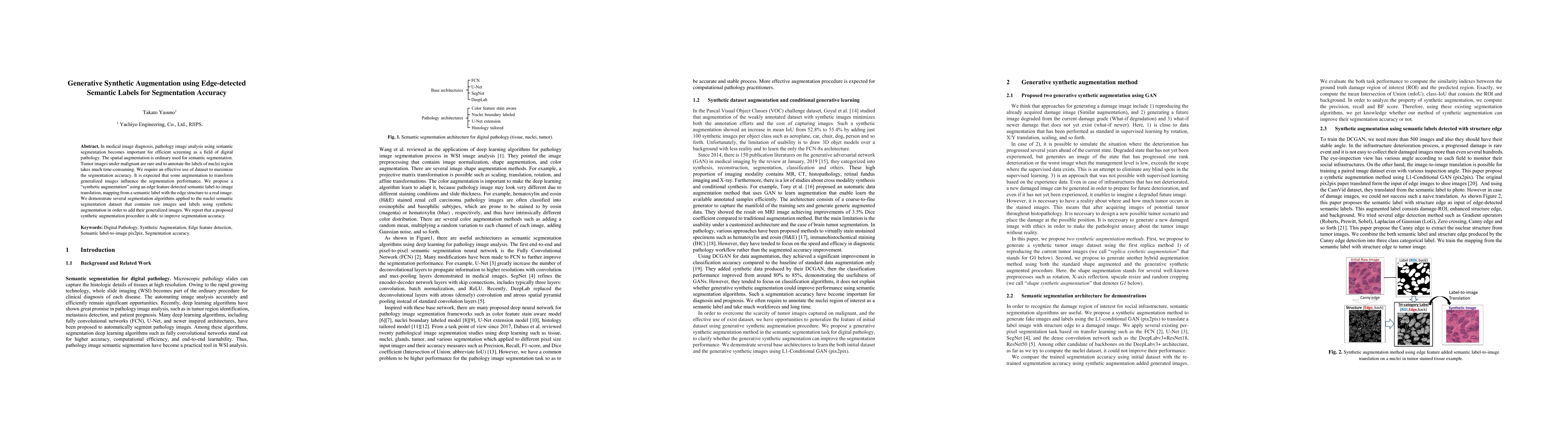
In medical image diagnosis, pathology image analysis using semantic segmentation becomes important for efficient screening as a field of digital pathology. The spatial augmentation is ordinary used for semantic segmentation. Tumor images under malignant are rare and to annotate the labels of nuclei region takes much time-consuming. We require an effective use of dataset to maximize the segmentation accuracy. It is expected that some augmentation to transform generalized images influence the segmentation performance. We propose a synthetic augmentation using label-to-image translation, mapping from a semantic label with the edge structure to a real image. Exactly this paper deal with stain slides of nuclei in tumor. Actually, we demonstrate several segmentation algorithms applied to the initial dataset that contains real images and labels using synthetic augmentation in order to add their generalized images. We computes and reports that a proposed synthetic augmentation procedure improve their accuracy.
AI Key Findings
Get AI-generated insights about this paper's methodology, results, significance, and more — seven facets brought into focus.
Impact
Paper Details
PDF Preview
Key Terms
Citation Network
Current paper (gray), citations (green), references (blue)
Display is limited for performance on very large graphs.
Discussion 0