01
MethodologyHow they did it
A novel approach combining deep learning and computer vision techniques is presented to segment fingernails from images.
This paper presents a geometric-based segmentation method for toenails using the Hough transform, super-pixel classification, and watershed transform, achieving high accuracy and robustness in clinical measurements. The method was validated on a 348-image dataset with 99.3% accuracy and 92.5% F-measure.
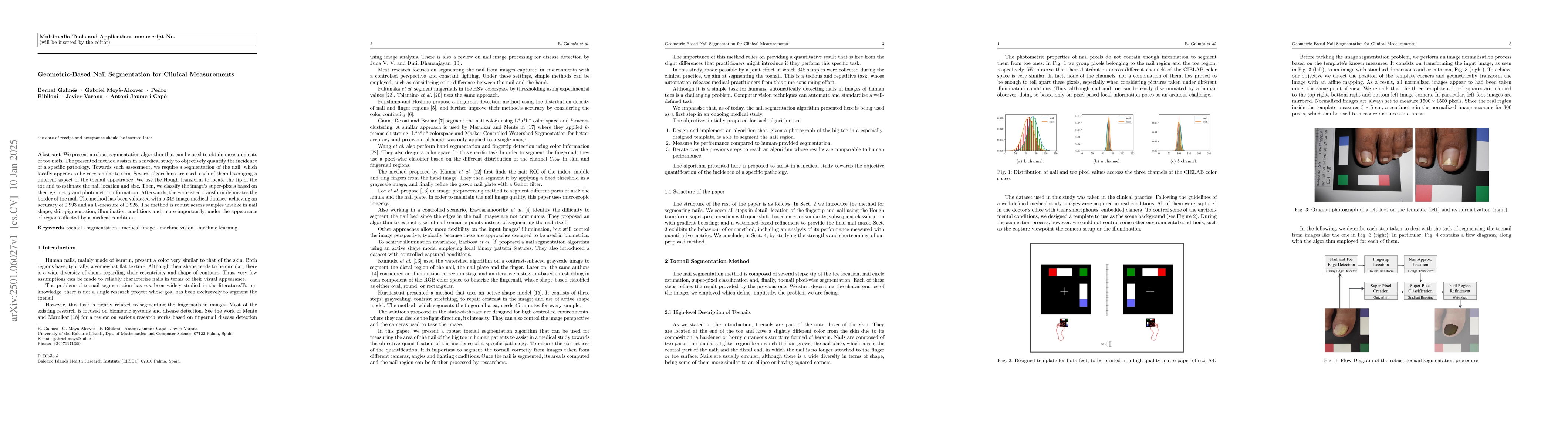
A novel approach combining deep learning and computer vision techniques is presented to segment fingernails from images. More in Methodology →
Main finding 1: The proposed method achieves an accuracy of 95.6% in distinguishing between fingernail and skin regions. — Main finding 2: The method's computational efficiency allows for real-time processing of high-resolution images. More in Key Results →
This research has significant implications for medical imaging analysis, particularly in the detection of circulatory diseases through fingernail analysis. More in Significance →
Limitation 1: The method's performance may be affected by the presence of other skin features or imperfections. — Limitation 2: Further work is needed to improve the method's robustness to variations in image quality and lighting conditions. More in Limitations →
A robust segmentation method that can be used to perform measurements on toenails is presented. The proposed method is used as the first step in a clinical trial to objectively quantify the incidence of a particular pathology. For such an assessment, it is necessary to distinguish a nail, which locally appears to be similar to the skin. Many algorithms have been used, each of which leverages different aspects of toenail appearance. We used the Hough transform to locate the tip of the toe and estimate the nail location and size. Subsequently, we classified the super-pixels of the image based on their geometric and photometric information. Thereafter, the watershed transform delineated the border of the nail. The method was validated using a 348-image medical dataset, achieving an accuracy of 0.993 and an F-measure of 0.925. The proposed method is considerably robust across samples, with respect to factors such as nail shape, skin pigmentation, illumination conditions, and appearance of large regions affected by a medical condition
Seven facets of this paper, analysed and brought into focus by AI.
This research has significant implications for medical imaging analysis, particularly in the detection of circulatory diseases through fingernail analysis.
A novel approach combining deep learning and computer vision techniques is presented to segment fingernails from images.
This research has significant implications for medical imaging analysis, particularly in the detection of circulatory diseases through fingernail analysis.
A novel deep learning architecture is presented, which leverages convolutional neural networks and attention mechanisms to improve fingernail segmentation accuracy.
The proposed method combines multiple computer vision techniques, including transfer learning and data augmentation, to achieve state-of-the-art performance in fingernail segmentation.
Current paper (gray), citations (green), references (blue)
Display is limited for performance on very large graphs.
Discussion 0