01
MethodologyHow they did it
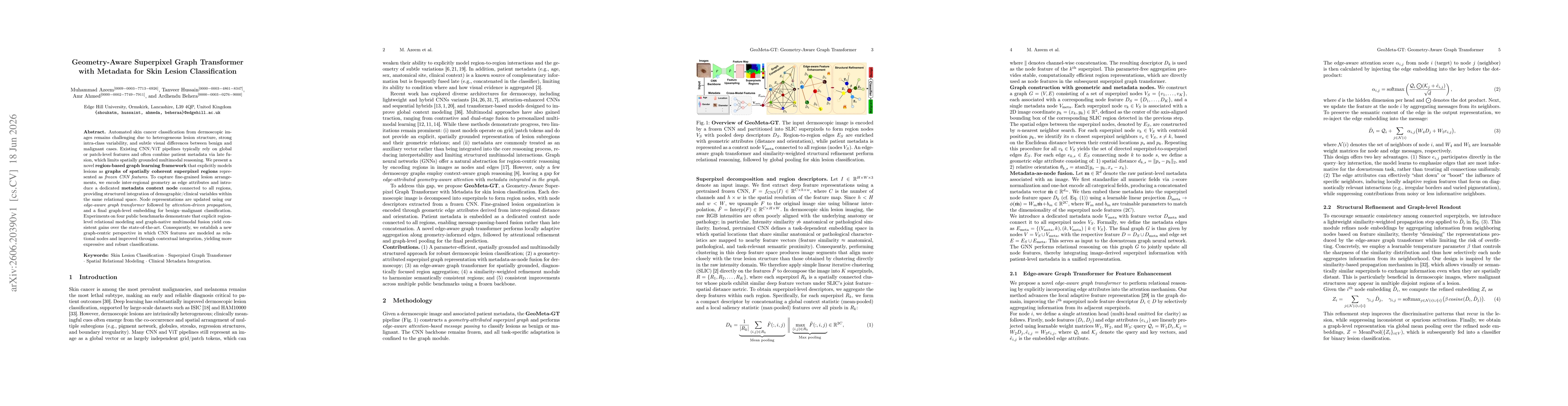
GeoMeta-GT constructs a geometry-aware, region-centric graph from dermoscopic images by decomposing each image into superpixels whose frozen CNN features form the region nodes. Inter-regional geometry is encoded as edge attributes, and a dedicated metadata context node connected to all regions enables structured fusion of demographic/clinical variables within the graph. An edge-aware graph transformer updates node representations via geometry-informed message passing, followed by attention-driven refinement and graph pooling for benign/malignant classification. The backbone CNN remains frozen and task-specific adaptations are confined to the graph module, emphasizing a graph-centric reasoning workflow with multimodal integration.
Discussion 0