Publication
Metrics
AI Quick Summary
This paper introduces the Geometrical Variation Method (GVM) for estimating stress within epithelial tissues by analyzing cellular geometry from images. The GVM leverages the balance of local line tension and pressure differentials to infer stress, providing a robust method for non-destructive stress estimation in live tissues.
Paper Preview
Abstract
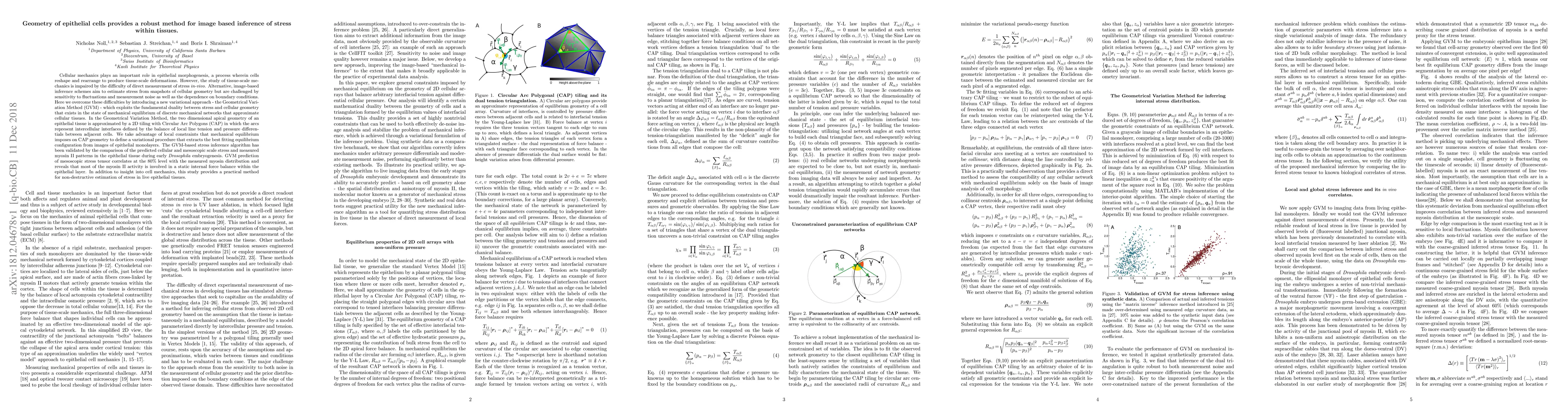
Cellular mechanics plays an important role in epithelial morphogenesis, a process wherein cells reshape and rearrange to produce tissue-scale deformations. However, the study of tissue-scale mechanics is impaired by the difficulty of direct measurement of stress in-vivo. Alternative, image-based inference schemes aim to estimate stress from snapshots of cellular geometry but are challenged by sensitivity to fluctuations and measurement noise as well as the dependence on boundary conditions. Here we overcome these difficulties by introducing a new variational approach - the Geometrical Variation Method (GVM) - which exploits the fundamental duality between stress and cellular geometry that exists in the state of mechanical equilibrium of discrete mechanical networks that approximate cellular tissues. In the Geometrical Variation Method, the two dimensional apical geometry of an epithelial tissue is approximated by a 2D tiling with Circular Arc Polygons (CAP) in which the arcs represent intercellular interfaces defined by the balance of local line tension and pressure differentials between adjacent cells. We take advantage of local constraints that mechanical equilibrium imposes on CAP geometry to define a variational procedure that extracts the best fitting equilibrium configuration from images of epithelial monolayers. The GVM-based stress inference algorithm has been validated by the comparison of the predicted cellular and mesoscopic scale stress and measured myosin II patterns in the epithelial tissue during Drosophila embryogenesis. GVM prediction of mesoscopic stress tensor correlates at the 80% level with the measured myosin distribution and reveals that most of the myosin II activity is involved in a static internal force balance within the epithelial layer. Lastly, this study provides a practical method for non-destructive estimation of stress in live epithelial tissues.
AI Key Findings
Get AI-generated insights about this paper's methodology, results, significance, and more — seven facets brought into focus.
Impact
Paper Details
Authors
PDF Preview
Key Terms
Citation Network
Current paper (gray), citations (green), references (blue)
Display is limited for performance on very large graphs.
Discussion 0