Quick Summary:

This paper introduces algorithms for automatic registration and 3D reconstruction of conventionally produced mouse brain slices, significantly improving the handling of histological artifacts. It employs geometric methods and non-linear registration to align slice images with the Allen Reference Atlas, enhancing neuron counting in anatomical regions.

Summary

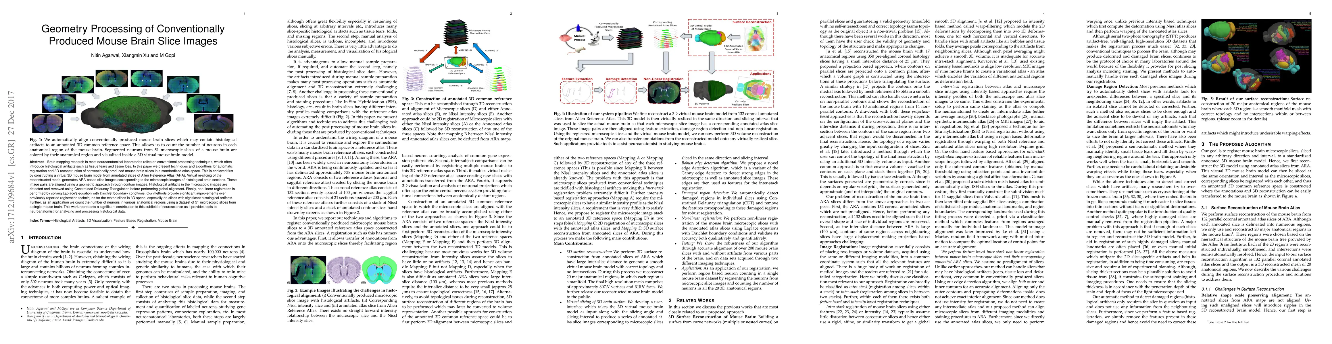
Brain mapping research in most neuroanatomical laboratories relies on conventional processing techniques, which often introduce histological artifacts such as tissue tears and tissue loss. In this paper we present techniques and algorithms for automatic registration and 3D reconstruction of conventionally produced mouse brain slices in a standardized atlas space. This is achieved first by constructing a virtual 3D mouse brain model from annotated slices of Allen Reference Atlas (ARA). Virtual re-slicing of the reconstructed model generates ARA-based slice images corresponding to the microscopic images of histological brain sections. These image pairs are aligned using a geometric approach through contour images. Histological artifacts in the microscopic images are detected and removed using Constrained Delaunay Triangulation before performing global alignment. Finally, non-linear registration is performed by solving Laplace's equation with Dirichlet boundary conditions. Our methods provide significant improvements over previously reported registration techniques for the tested slices in 3D space, especially on slices with significant histological artifacts. Further, as an application we count the number of neurons in various anatomical regions using a dataset of 51 microscopic slices from a single mouse brain. This work represents a significant contribution to this subfield of neuroscience as it provides tools to neuroanatomist for analyzing and processing histological data.

AI Key Findings

Get AI-generated insights about this paper's methodology, results, and significance.

Paper Details

PDF Preview

Download PDF

Key Terms

brain (0.315)
artifacts (0.264)
microscopic (0.224)
atlas (0.219)
images (0.196)
virtual (0.192)
techniques (0.163)
processing (0.157)
significant (0.154)
neurons (0.134)
laplace (0.124)
dirichlet (0.123)
removed (0.123)
count (0.122)
reconstructed (0.122)
annotated (0.121)
aligned (0.116)
represents (0.113)
alignment (0.110)
performing (0.109)

Citation Network

Current paper (gray), citations (green), references (blue)

Current Paper
Citations
References
Click to view
Title Authors Year Actions
RegBoost: Enhancing mouse brain image registration using geometric priors and Laplacian interpolation
Atchuth Naveen Chilaparasetti, Andy Thai, Pan Gao, Xiangmin Xu, M. Gopi
2024
View Paper
Molecular signatures of cortical expansion in the human foetal brain
G. Ball, S. Oldham, V. Kyriakopoulou, L. Z. Williams, V. Karolis, A. Price, J. Hutter, M. Seal, A. Alexander-Bloch, J. Hajnal, A. Edwards, E. C. Robinson, J. Seidlitz
2024
View Paper
Robotic Patch Clamp Electrophysiological Signal Measurement in Multiple Brain Regions Based on Brain Slice Registration
Ke Li, Huiying Gong, Jinyu Qiu, Ruimin Li, Qili Zhao, Xin Zhao, Mingzhu Sun
2024
View Paper
Fine-tuning TrailMap: The utility of transfer learning to improve the performance of deep learning in axon segmentation of light-sheet microscopy images
Marjolein Oostrom, Michael A. Muniak, Rogene Eichler West, Sarah Akers, Paritosh Pande, Moses Obiri, Wei Wang, Kasey Bowyer, Zhuhao Wu, Lisa M. Bramer, Tianyi Mao, B. Webb-Robertson
2024
View Paper
3-Dimensional Reconstruction from Histopathological Sections: A Systematic Review.
Alexander Kurz, Heimo Müller, J. N. Kather, Lucas Schneider, Tabea-Clara Bucher, T. Brinker
2024
View Paper
Molecular signatures of cortical expansion in the human fetal brain
G. Ball, S. Oldham, V. Kyriakopoulou, L. Z. Williams, V. Karolis, A. Price, J. Hutter, M. Seal, A. Alexander-Bloch, J. Hajnal, A. Edwards, E. C. Robinson, J. Seidlitz
2024
View Paper
Automated identification of protein expression intensity and classification of protein cellular locations in mouse brain regions from immunofluorescence images
Lin-Xia Bao, Zhuo-Ming Luo, Xi-Liang Zhu, Ying-Ying Xu
2023
View Paper
DLATA: Deep Learning-Assisted Transformation Alignment of 2D Brain Slice Histology
Moxuan Luo, Qingqing Liu, Liping Wang, Rosa H. M. Chan
2023
View Paper
Expected affine: A registration method for damaged section in serial sections electron microscopy
T. Xin, Lijun Shen, Linlin Li, Xi Chen, Hua Han
2022
View Paper
BoutonNet: an automatic method to detect anterogradely labeled presynaptic boutons in brain tissue sections
Fillan Grady, Shantelle A. Graff, Georgina M. Aldridge, Joel C Geerling
2022
View Paper
Automatic 2D to 3D localization of histological mouse brain sections in the reference atlas using deep learning
Maryam Sadeghi, Pedro Neto, Arnau Ramos-Prats, Federico Castaldi, Enrica Paradiso, N. Mahmoodian, F. Ferraguti, Georg Goebel
2022
View Paper
Slice2Volume: Fusion of Multimodal Medical Imaging and Light Microscopy Data of Irradiation-Injured Brain Tissue in 3D.
Soltwedel Johannes, Suckert Theresa, Beyreuther Elke, Schneider Moritz, Boucsein Marc, Bodenstein Elisabeth, Nexhipi Sindi, Stolz-Kieslich Liane, Krause Mechthild, von Neubeck Cläre, Haase Robert, Lühr Armin, Dietrich Antje
2021
View Paper
A simple method defines 3D morphology and axon projections of filled neurons in a small CNS volume: Steps toward understanding functional network circuitry
D. Conte, R. Borisyuk, M. Hull, A. Roberts
2020
View Paper
3D Mapping of Serial Histology Sections with Anomalies Using a Novel Robust Deformable Registration Algorithm
D. Tward, Xu Li, Bingxing Huo, Brian C. Lee, P. Mitra, M. Miller
2019
View Paper
Stacked in-plane histology for quantitative validation of non-invasive imaging biomarkers: Application to an infiltrative brain tumour model
H. Al-Mubarak, A. Vallatos, L. Gallagher, J. Birch, L. Gilmour, J. Foster, A. Chalmers, W. Holmes
2019
View Paper
Detection of neural connections with ex vivo MRI using a ferritin-encoding trans-synaptic virus
Ning Zheng, Peng Su, Yue Liu, Huadong Wang, B. Nie, Xiaohui Fang, Yue Xu, Kunzhang Lin, Pei Lv, Xiaobin He, Yi Guo, B. Shan, A. Manyande, Jie Wang, Fuqiang Xu
2019
View Paper
ARMBIS: accurate and robust matching of brain image sequences from multiple modal imaging techniques
Qi Shen, Goayu Xiao, Yingwei Zheng, Jie Wang, Yue Liu, Xutao Zhu, Fan Jia, Peng Su, B. Nie, Fuqiang Xu, Bin Zhang
2019
View Paper
Exploiting Gene Expression Profiles for the Automated Prediction of Connectivity between Brain Regions
I. Roberti, Marta Lovino, Santa Di Cataldo, E. Ficarra, Gianvito Urgese
2019
View Paper

Comments (0)

This website uses cookies to ensure you get the best experience. Learn more