This paper proposes a novel deep learning framework called deep multichannel side supervision (DMCS) for segmenting individual glands in colon histology images. The method leverages convolutional neural networks and deep supervision to automatically extract and fuse complex multichannel information, achieving state-of-the-art results compared to previous methods.
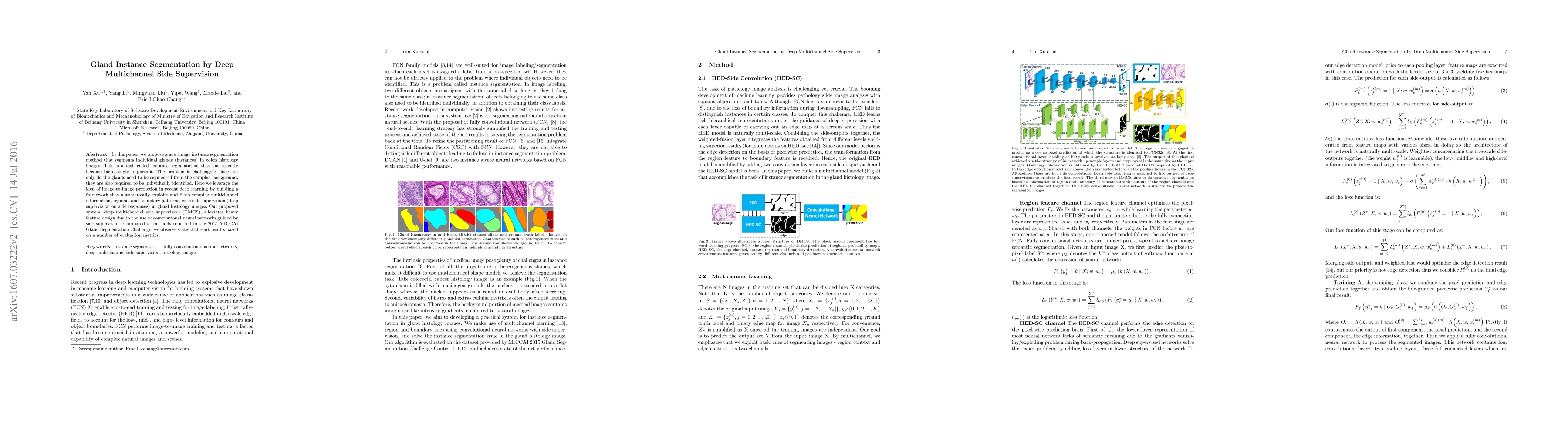
In this paper, we propose a new image instance segmentation method that segments individual glands (instances) in colon histology images. This is a task called instance segmentation that has recently become increasingly important. The problem is challenging since not only do the glands need to be segmented from the complex background, they are also required to be individually identified. Here we leverage the idea of image-to-image prediction in recent deep learning by building a framework that automatically exploits and fuses complex multichannel information, regional and boundary patterns, with side supervision (deep supervision on side responses) in gland histology images. Our proposed system, deep multichannel side supervision (DMCS), alleviates heavy feature design due to the use of convolutional neural networks guided by side supervision. Compared to methods reported in the 2015 MICCAI Gland Segmentation Challenge, we observe state-of-the-art results based on a number of evaluation metrics.
This graph shows how this paper is connected to other research. The current paper is shown in dark gray, papers that cite it are in green, and papers it references are in blue. You can:
• Hover over any node to see the paper title
• Click a node to view that paper's details
• Drag nodes to rearrange the graph
• Use the zoom controls (bottom right) to zoom in/out
Title | Authors | Year | Actions |
---|---|---|---|
Graph confidence intercalibration network for intracranial aneurysm lesion instance segmentation in DSA
|
Haili Ye, Yancheng Mo, Chen Tang, Mingqian Liao, Xiaoqing Zhang, Limeng Dai, Baihua Li, Jiang Liu
|
2024
|
View Paper |
MaskUno: Switch-Split Block For Enhancing Instance Segmentation
|
Jawad Haidar, Marc Mouawad, Imad H. Elhajj, Daniel Asmar
|
2024
|
View Paper |
Contour and Enclosed Region Refining for Contour-Based Instance Segmentation
|
Wenchao Gu, Shuang Bai
|
2023
|
View Paper |
MLAGG-Net: Multi-level aggregation and global guidance network for pancreatic lesion segmentation in histopathological images
|
Ao Liu, Huiyan Jiang, Weiwei Cao, Wenju Cui, Dehui Xiang, C. Shao, Zhaobang Liu, Yun Bian, Jian Zheng
|
2023
|
View Paper |
DenseRes-Net: An Architecture for Gland Segmentation
|
Wangkheirakpam Reema Devi, S. Roy, Khelchandra Thongam, Chiranjiv Chingangbam
|
2023
|
View Paper |
DARMF-UNet: A dual-branch attention-guided refinement network with multi-scale features fusion U-Net for gland segmentation
|
Junmei Sun, Xin Zhang, Xiumei Li, Ruyu Liu, Tianyang Wang
|
2023
|
View Paper |
Dense Dilated Multi-Scale Supervised Attention-Guided Network for histopathology image segmentation
|
Rangan Das, Shirsha Bose, Ritesh Sur Chowdhury, U. Maulik
|
2023
|
View Paper |
On the Analyses of Medical Images Using Traditional Machine Learning Techniques and Convolutional Neural Networks
|
Saeed Iqbal, Adnan N. Qureshi, Jianqiang Li, Tariq Mahmood
|
2023
|
View Paper |
An Atrous Convolved Hybrid Seg-Net Model with residual and attention mechanism for gland detection and segmentation in histopathological images
|
Manju Dabass, Jyoti Dabass
|
2023
|
View Paper |
Automatic multi-tissue segmentation in pancreatic pathological images with selected multi-scale attention network
|
Enting Gao, Huiyan Jiang, Zhibang Zhou, Changxing Yang, Muyang Chen, Weifang Zhu, Fei Shi, Xinjian Chen, Jian Zheng, Yun Bian, Dehui Xiang
|
2022
|
View Paper |
Two Path Gland Segmentation Algorithm of Colon Pathological Image Based on Local Semantic Guidance
|
Songtao Ding, Hongyu Wang, Hu Lu, M. Nappi, Shaohua Wan
|
2022
|
View Paper |
GOAT: Deep learning-enhanced Generalized Organoid Annotation Tool
|
Jan P. Bremer, M. Baumdick, Marius S. Knorr, Lucy Wegner, Jasmin Wesche, A. Jordan-Paiz, Johannes M. Jung, A. Highton, Julia Jäger, Ole Hinrichs, Sébastien Brias, Jennifer Niersch, L. Müller, R. R. Schreurs, T. Koyro, Sebastian M. Löbl, Leonore Mensching, L. Konczalla, Annika Niehrs, F. Vondran, Christoph Schramm, Angelique Hölzemer, K. Oldhafer, I. Königs, Stefan Kluge, D. Perez, K. Reinshagen, S. Pals, N. Gagliani, Sander Joosten, M. Topf, M. Altfeld, M. Bunders
|
2022
|
View Paper |
BE-Net:Boudary-Enhanced Networks for Accurate Gland Segmentation
|
Zhenbang Fan, Sheng Dong, Shuling Shi, Wenqin Yang, Sheng Li, Xiongxiong He
|
2022
|
View Paper |
Divide-and-Attention Network for HE-Stained Pathological Image Classification
|
Rui Yan, Zhidong Yang, Jintao Li, Chunhou Zheng, Fa Zhang
|
2022
|
View Paper |
Nuclei-Guided Network for Breast Cancer Grading in HE-Stained Pathological Images
|
Rui Yan, Fei Ren, Jintao Li, Xiaosong Rao, Zhilong Lv, Chunhou Zheng, Fa Zhang
|
2022
|
View Paper |
An application for wound diagnosis and treatment
|
Lázaro Gilvan Rodrigues da Silva, Anderson Aparecido Alves da Silva, A. Guelfi, M. N. de Rezende, Jose de Jesus Perez Alcazar, Marcelo Teixeira de Azevedo, S. Kofuji
|
2022
|
View Paper |
Artificial Intelligence Program to Predict p53 Mutations in Ulcerative Colitis-Associated Cancer or Dysplasia.
|
T. Noguchi, Takumi Ando, S. Emoto, H. Nozawa, K. Kawai, K. Sasaki, K. Murono, J. Kishikawa, Hiroaki Ishi, Y. Yokoyama, S. Abe, Yuzo Nagai, H. Anzai, H. Sonoda, K. Hata, T. Sasaki, S. Ishihara
|
2022
|
View Paper |
MRI image analysis for cerebrum tumor detection and feature extraction using 2D U-ConvNet and SVM classification
|
R. Pitchai, P. Supraja, A. Sulthana, T. Veeramakali, Christy M Babu
|
2022
|
View Paper |
A review on 2D instance segmentation based on deep neural networks
|
Wenchao Gu, Shuang Bai, Ling-Yun Kong
|
2022
|
View Paper |
Boosting Boundary Representation for Gland Instance Segmentation
|
Yuxin Kang, Hansheng Li, Zhuoyue Wu, Feihong Liu, D. Hu, Lei Cui, Jun Feng, Lin Yang
|
2021
|
View Paper |
Real-time Instance Segmentation of Surgical Instruments using Attention and Multi-scale Feature Fusion
|
Juan Carlos Angeles Ceron, Gilberto Ochoa-Ruiz, Leonardo Chang, Sharib Ali
|
2021
|
View Paper |
Fusion Coding of 3D Real and Virtual Scenes Information for Augmented Reality-Based Holographic Stereogram
|
Yunpeng Liu, Xingpeng Yan, Xinlei Liu, Xi Wang, Tao Jing, Min Lin, Song Chen, Pei-pei Li, Xiaoyu Jiang
|
2021
|
View Paper |
Automated Gland Detection in Colorectal Histopathological Images
|
Maisun Mohamed Al Zorgani, I. Mehmood, H. Ugail
|
2021
|
View Paper |
Deep Learning Applications in Medical Image Analysis
|
A. Singha, R. Thakur, Tushar Patel
|
2021
|
View Paper |
The future of bone regeneration: integrating AI into tissue engineering
|
Benita Scout Mackay, Karen Marshall, J. Grant-Jacob, J. Kanczler, R. Eason, R. Oreffo, B. Mills
|
2021
|
View Paper |
Glandular Cell Segmentation Method Combined with Dense Connective Blocks and Self-Attention Mechanism
|
Baoqi Zhao, Fei Yu, Junmei Sun, Xiumei Li, L. Yuan, Lei Xiao
|
2021
|
View Paper |
Learning Melanocytic Proliferation Segmentation in Histopathology Images from Imperfect Annotations
|
Kechun Liu, M. Mokhtari, Beibin Li, Shima Nofallah, Caitlin J. May, Oliver H. Chang, S. Knezevich, J. Elmore, Linda G. Shapiro
|
2021
|
View Paper |
State of machine and deep learning in histopathological applications in digestive diseases
|
Soma Kobayashi, J. Saltz, V. Yang
|
2021
|
View Paper |
Weakly-Supervised Gland Instance Segmentation based on Point Labeling
|
Da-han Wang, Haili Ye, Jianmin Li, Shunzhi Zhu, Chenyan Zhu
|
2021
|
View Paper |
Every Annotation Counts: Multi-label Deep Supervision for Medical Image Segmentation
|
Simon Reiß, Constantin Seibold, Alexander Freytag, E. Rodner, R. Stiefelhagen
|
2021
|
View Paper |
Artificial intelligence and digital pathology: Opportunities and implications for immuno-oncology
|
F. Sobhani, R. Robinson, A. Hamidinekoo, I. Roxanis, N. Somaiah, Yinyin Yuan
|
2021
|
View Paper |
LR-Net: A Multi-task Model Using Relationship-based Contour Information to Enhance the Semantic Segmentation of Cancer Regions
|
Baorong Shi, Hong Zhang, Rui Yan, Wang Jing, Jinfeng Zang, Fa Zhang
|
2020
|
View Paper |
Constrained Multi-scale Dense Connections for Accurate Biomedical Image Segmentation
|
Jiawei Zhang, Yanchun Zhang, Shanfeng Zhu, Xiaowei Xu
|
2020
|
View Paper |
Maximal Information Complemented Refinement Network for Gland Instance Segmentation
|
Bowen Xu, Yankai Wang, Dawei Yang, Wei Zhang, Ye Zhang, Yajie Kong, Wenqiang Zhang
|
2020
|
View Paper |
NANet: Nuclei-Aware Network for Grading of Breast Cancer in HE Stained Pathological Images
|
Rui Yan, Jintao Li, Xiaosong Rao, Zhilong Lv, Chunhou Zheng, Jinjin Dou, Xiaowen Wang, Fei Ren, Fa Zhang
|
2020
|
View Paper |
Cerebrum Tumor Segmentation of High Resolution Magnetic Resonance Images Using 2D-Convolutional Network with Skull Stripping
|
R. Pitchai, Ch Madhu Babu, P. Supraja, Mahesh Kumar Challa
|
2020
|
View Paper |
3D convolutional neural networks-based segmentation to acquire quantitative criteria of the nucleus during mouse embryogenesis
|
Y. Tokuoka, Takahiro G. Yamada, Daisuke Mashiko, Zenki Ikeda, N. Hiroi, Tetsuya J. Kobayashi, Kazuo Yamagata, Akira Funahashi
|
2020
|
View Paper |
A Simultaneous Object Detection and Component Segmentation Approach Based on Mask R-CNN
|
Deshuai Zheng, Jin Yan, Yong Liu, Tao Xue
|
2020
|
View Paper |
Artificial Intelligence based analysis to evaluate for RBC cells, Pus cells and Calcium Oxalate crystals in Urine sediment slide microscopy: A pilot study in North India
|
Ankita Agrawal, Shelley Saxena
|
2020
|
View Paper |
AttentionBoost: Learning What to Attend for Gland Segmentation in Histopathological Images by Boosting Fully Convolutional Networks
|
G. N. Gunesli, C. Sokmensuer, C. Gunduz-Demir
|
2020
|
View Paper |
A Stacked Generalization U-shape network based on zoom strategy and its application in biomedical image segmentation
|
Tianyu Shi, Huiyan Jiang, Bin Zheng
|
2020
|
View Paper |
LevelSet R-CNN: A Deep Variational Method for Instance Segmentation
|
N. Homayounfar, Yuwen Xiong, Justin Liang, Wei-Chiu Ma, R. Urtasun
|
2020
|
View Paper |
Deep learning in digital pathology image analysis: a survey
|
Shujian Deng, Xin Zhang, Wen Yan, E. Chang, Yubo Fan, M. Lai, Yan Xu
|
2020
|
View Paper |
Multiple Morphological Constraints-Based Complex Gland Segmentation in Colorectal Cancer Pathology Image Analysis
|
Kun Zhang, JunHong Fu, Liang Hua, Peijian Zhang, Y. Shao, Sheng Xu, Huiyu Zhou, L. Chen, Jing Wang
|
2020
|
View Paper |
On uncertainty estimation in active learning for image segmentation
|
Bo Li, T. S. Alstrøm
|
2020
|
View Paper |
SCAU-Net: Spatial-Channel Attention U-Net for Gland Segmentation
|
P. Zhao, Jindi Zhang, W. Fang, Shuiguang Deng
|
2020
|
View Paper |
Dense Contour-Imbalance Aware framework for Colon Gland Instance Segmentation
|
Liye Mei, Xiaopeng Guo, Xin Huang, Yueyun Weng, Sheng Liu, Cheng Lei
|
2020
|
View Paper |
Deep Learning Based Accurate Hepatic Steatosis Quantification for Histological Assessment of Liver Biopsies
|
M. Roy, Fusheng Wang, Hoang Vo, Dejun Teng, George Teodoro, A. Farris, Eduardo Castillo‐Leon, M. Vos, Jun Kong
|
2020
|
View Paper |
Segmentation of Cell Images Based on Improved Deep Learning Approach
|
Chuanbo Huang, Huali Ding, Chuanling Liu
|
2020
|
View Paper |
Poorly Differentiated Colorectal Gland Segmentation Approach Based on Internal and External Stress in Histology Images
|
JunHong Fu, Kun Zhang, Peijian Zhang
|
2020
|
View Paper |
To evaluate AI for Hypochromic Anaemia based on RBC morphology in Leishman stained blood smears: A pilot study
|
R. Kushwaha, Ankit Agarwal, Shelley Saxena
|
2020
|
View Paper |
OR-Skip-Net: Outer residual skip network for skin segmentation in non-ideal situations
|
Muhammad Arsalan, Dong Seop Kim, Muhammad Owais, K. Park
|
2020
|
View Paper |
Automatic Polyp Recognition in Colonoscopy Images Using Deep Learning and Two-Stage Pyramidal Feature Prediction
|
Xiao Jia, Xiaochun Mai, Yi Cui, Yixuan Yuan, Xiaohan Xing, Hyunseok Seo, L. Xing, M. Meng
|
2020
|
View Paper |
Enabling a Single Deep Learning Model for Accurate Gland Instance Segmentation: A Shape-Aware Adversarial Learning Framework
|
Zengqiang Yan, Xin Yang, Kwang-Ting Cheng
|
2020
|
View Paper |
Wireless Capsule Endoscopy: A New Tool for Cancer Screening in the Colon With Deep-Learning-Based Polyp Recognition
|
Xiao Jia, Xiaohan Xing, Yixuan Yuan, L. Xing, M. Meng
|
2020
|
View Paper |
Deep neural network models for computational histopathology: A survey
|
C. Srinidhi, Ozan Ciga, Anne L. Martel
|
2019
|
View Paper |
Artificial Intelligence based AFB microscopy for Pulmonary Tuberculosis in North India: A pilot study
|
V. Khare, Ankita Agrawal, Prashant Gupta, Shelley Saxena
|
2019
|
View Paper |
Hematoxylin and Eosin (H&E) Stained Liver Portal Area Segmentation Using Multi-Scale Receptive Field Convolutional Neural Network
|
Qi-En Xiao, P. Chung, H. Tsai, K. Cheng, N. Chow, Y. Juang, H. Tsai, Cheng-Hsiung Wang, Tsan-An Hsieh
|
2019
|
View Paper |
DPANet: A Novel Network Based on Dense Pyramid Feature Extractor and Dual Correlation Analysis Attention Modules for Colon Glands Segmentation
|
Shuting Liu, Baochang Zhang, Xi Li, Yiqing Liu, Mengying Hu, Tian Guan, Yonghong He
|
2019
|
View Paper |
Improving Nuclei/Gland Instance Segmentation in Histopathology Images by Full Resolution Neural Network and Spatial Constrained Loss
|
Hui Qu, Zhennan Yan, Gregory M. Riedlinger, Subhajyoti De, Dimitris N. Metaxas
|
2019
|
View Paper |
AttentionBoost: Learning What to Attend by Boosting Fully Convolutional Networks
|
G. N. Gunesli, C. Sokmensuer, Cigdem Demir
|
2019
|
View Paper |
Multi-Organ Gland Segmentation Using Deep Learning
|
T. Binder, El Mehdi Tantaoui, Pushpak Pati, R. Catena, Ago Set-Aghayan, M. Gabrani
|
2019
|
View Paper |
An Effective Cell Pathology Image Detection Method Based on Deep Stacked Auto-Encoder Combined with Random Forest
|
Xin Yan, Lei Wang
|
2019
|
View Paper |
Gland Segmentation in Histopathology Images Using Deep Networks and Handcrafted Features
|
Safiyeh Rezaei, Ali Emami, Hamidreza Zarrabi, Shima Rafiei, K. Najarian, N. Karimi, S. Samavi, S. Soroushmehr
|
2019
|
View Paper |
Automated segmentation of cell membranes to evaluate HER2 status in whole slide images using a modified deep learning network
|
Fariba D. Khameneh, Salar Razavi, M. Kamasak
|
2019
|
View Paper |
A New Framework to Reduce Doctor’s Workload for Medical Image Annotation
|
Yang Deng, Yao Sun, Yongpei Zhu, Yue Xu, Qianxi Yang, Shuo Zhang, Zhanyu Wang, Jirang Sun, Weiling Zhao, Xiaobo Zhou, Kehong Yuan
|
2019
|
View Paper |
The Classification of Renal Cancer in 3-Phase CT Images Using a Deep Learning Method
|
Seokmin Han, S. Hwang, H. Lee
|
2019
|
View Paper |
Adaptive Weighting Multi-Field-Of-View CNN for Semantic Segmentation in Pathology
|
Hiroki Tokunaga, Y. Teramoto, A. Yoshizawa, Ryoma Bise
|
2019
|
View Paper |
Artificial intelligence for microscopy: what you should know.
|
Lucas von Chamier, Romain F. Laine, Ricardo Henriques
|
2019
|
View Paper |
Efforts estimation of doctors annotating medical image
|
Yang Deng, Yao Sun, Yongpei Zhu, Yue Xu, Qianxi Yang, Shuo Zhang, Mingwang Zhu, Jirang Sun, Weiling Zhao, Xiaobo Zhou, Kehong Yuan
|
2019
|
View Paper |
SPDA: Superpixel-based Data Augmentation for Biomedical Image Segmentation
|
Yizhe Zhang, Lin Yang, Hao Zheng, Peixian Liang, Colleen A. Mangold, R. Loreto, David P. Hughes, D. Chen
|
2018
|
View Paper |
Tissue Region Growing for Hispathology Image Segmentation
|
Zhaoyang Xu, C. F. Moro, Danyil Kuznyecov, B. Bozóky, Le Dong, Qianni Zhang
|
2018
|
View Paper |
Automatic segmentation of esophageal cancer pathological sections based on semantic segmentation
|
Sizhe Li, Jiawei Zhang, Yuzhen Jin, Liping Zheng, Jilan Xu, Guanzhen Yu, Yanchun Zhang
|
2018
|
View Paper |
Fusion of encoder-decoder deep networks improves delineation of multiple nuclear phenotypes
|
M. Khoshdeli, G. Winkelmaier, B. Parvin
|
2018
|
View Paper |
Transfer Learning and Data Augmentation for Semantic Segmentation in Histopathology
|
Giorgia Cantisani
|
2018
|
View Paper |
MILD‐Net: Minimal information loss dilated network for gland instance segmentation in colon histology images
|
S. Graham, Hao Chen, Q. Dou, P. Heng, N. Rajpoot
|
2018
|
View Paper |
Micro‐Net: A unified model for segmentation of various objects in microscopy images
|
S. Raza, Linda Cheung, M. Shaban, S. Graham, David B. A. Epstein, S. Pelengaris, Michael Khan, N. Rajpoot
|
2018
|
View Paper |
In Silico Labeling: Predicting Fluorescent Labels in Unlabeled Images
|
Eric M. Christiansen, Samuel J. Yang, D. Ando, A. Javaherian, G. Skibinski, S. Lipnick, Elliot Mount, Alison O’Neil, K. Shah, Alicia K. Lee, P. Goyal, W. Fedus, R. Poplin, A. Esteva, Marc Berndl, Lee L. Rubin, Philip Nelson, S. Finkbeiner
|
2018
|
View Paper |
Quantization of Fully Convolutional Networks for Accurate Biomedical Image Segmentation
|
Xiaowei Xu, Q. Lu, Yu Hu, Lin Yang, X. Hu, D. Chen, Yiyu Shi
|
2018
|
View Paper |
Deep Deconvolutional Neural Network for Target Segmentation of Nasopharyngeal Cancer in Planning Computed Tomography Images
|
K. Men, Xinyuan Chen, Ye Zhang, Zhang Tao, J. Dai, J. Yi, Yexiong Li
|
2017
|
View Paper |
Automatic segmentation of the clinical target volume and organs at risk in the planning CT for rectal cancer using deep dilated convolutional neural networks
|
K. Men, J. Dai, Ye‐Xiong Li
|
2017
|
View Paper |
Segmentation and classification of colon glands with deep convolutional neural networks and total variation regularization
|
Philipp Kainz, Michael Pfeiffer, M. Urschler
|
2017
|
View Paper |
A deep learning framework for supporting the classification of breast lesions in ultrasound images
|
Seokmin Han, Hokyung Kang, J. Jeong, M. Park, Wonsik Kim, Won-Chul Bang, Y. Seong
|
2017
|
View Paper |
A Web-Based Platform for Distributed Annotation of Computerized Tomography Scans
|
N. Heller, P. Stanitsas, V. Morellas, N. Papanikolopoulos
|
2017
|
View Paper |
Boundary-Aware Fully Convolutional Network for Brain Tumor Segmentation
|
Haocheng Shen, Ruixuan Wang, Jianguo Zhang, S. McKenna
|
2017
|
View Paper |
Deep Adversarial Networks for Biomedical Image Segmentation Utilizing Unannotated Images
|
Yizhe Zhang, L. Yang, Jianxu Chen, Maridel Fredericksen, David P. Hughes, D. Chen
|
2017
|
View Paper |
Recent Advances in the Applications of Convolutional Neural Networks to Medical Image Contour Detection
|
Zizhao Zhang, Fuyong Xing, H. Su, Xiaoshuang Shi, L. Yang
|
2017
|
View Paper |
MIMONet: Gland Segmentation Using Multi-Input-Multi-Output Convolutional Neural Network
|
S. Raza, Linda Cheung, David B. A. Epstein, S. Pelengaris, Michael Khan, N. Rajpoot
|
2017
|
View Paper |
Suggestive Annotation: A Deep Active Learning Framework for Biomedical Image Segmentation
|
L. Yang, Yizhe Zhang, Jianxu Chen, Siyuan Zhang, D. Chen
|
2017
|
View Paper |
Stacked fully convolutional networks with multi-channel learning: application to medical image segmentation
|
Lei Bi, Jinman Kim, Ashnil Kumar, M. Fulham, D. Feng
|
2017
|
View Paper |
A survey on deep learning in medical image analysis
|
G. Litjens, Thijs Kooi, B. Bejnordi, A. Setio, F. Ciompi, Mohsen Ghafoorian, J. Laak, B. Ginneken, C. I. Sánchez
|
2017
|
View Paper |
DCAN: Deep contour‐aware networks for object instance segmentation from histology images
|
Hao Chen, Xiaojuan Qi, Lequan Yu, Q. Dou, J. Qin, P. Heng
|
2017
|
View Paper |
Gland Instance Segmentation Using Deep Multichannel Neural Networks
|
Yan Xu, Yang Li, Yipei Wang, Mingyuan Liu, Yubo Fan, M. Lai, E. Chang
|
2016
|
View Paper |
Gland Instance Segmentation by Deep Multichannel Neural Networks
|
Yan Xu, Y. Li, Mingyuan Liu, Yipei Wang, Yubo Fan, M. Lai, E. Chang
|
2016
|
View Paper |
UJAT-Net: A U-Net Combined Joint-Attention and Transformer for Breast Tubule Segmentation in H&E Stained Images
|
Li-Wang Liu, Zhao Huang, Kao-Yan Lu, Zu-Xuan Wang, Yao-Ming Liang, Shi-Yu Lin, Yan-Hong Ji
|
2024
|
View Paper |
ECAU-Net: Efficient channel attention U-Net for fetal ultrasound cerebellum segmentation
|
Xin Shu, Feng-Chen Chang, Xin Zhang, Changbin Shao, Xibei Yang
|
2022
|
View Paper |
From Pixel to Whole Slide: Automatic Detection of Microvascular Invasion in Hepatocellular Carcinoma on Histopathological Image via Cascaded Networks
|
Hanbo Chen, Kang Wang, Yuyao Zhu, Jiangpeng Yan, Yan Ji, Jingjing Li, D. Xie, Junzhou Huang, Shuqun Cheng, Jianhua Yao
|
2021
|
View Paper |
Attention-Guided deep atrous-residual U-Net architecture for automated gland segmentation in colon histopathology images
|
Manju Dabass, Sharda Vashisth, R. Vig
|
2021
|
View Paper |
High-Resolution Encoder–Decoder Networks for Low-Contrast Medical Image Segmentation
|
Sihang Zhou, Dong Nie, E. Adeli, Jianping Yin, J. Lian, D. Shen
|
2020
|
View Paper |
DoubleU-Net: Colorectal Cancer Diagnosis and Gland Instance Segmentation with Text-Guided Feature Control
|
Pei Wang, Albert C. S. Chung
|
2020
|
View Paper |
Recent Advances in Deep Learning Techniques and Its Applications: An Overview
|
A. Hazra, P. Choudhary, M. S. Singh
|
2020
|
View Paper |
Learning Hierarchical Semantic Correspondence and Gland Instance Segmentation
|
Pei Wang, Albert C. S. Chung
|
2020
|
View Paper |
Comprehensive Review on Deep Learning for Neuronal Disorders: Applications of Deep Learning
|
Vinayak Majhi, Angana Saikia, A. Datta, Aseem Sinha, S. Paul
|
2020
|
View Paper |
Multimodal medical volumes translation and segmentation with generative adversarial network
|
Zizhao Zhang, Lin Yang, Yefeng Zheng
|
2020
|
View Paper |
Deep Learning with Convolutional Neural Networks for Histopathology Image Analysis
|
D. Bosnacki, N. Riel, M. Veta
|
2019
|
View Paper |
Structure Prediction for Gland Segmentation With Hand-Crafted and Deep Convolutional Features
|
Siyamalan Manivannan, Wenqi Li, Jianguo Zhang, E. Trucco, S. McKenna
|
2018
|
View Paper |
Melanoma Diagnostics Using Fully Convolutional Networks on Whole Slide Images
|
Adon Phillips
|
2017
|
View Paper |
University of Dundee Boundary-aware fully convolutional network for brain tumor segmentation
|
Haocheng Shen
|
2017
|
View Paper |
World Journal of Gastroenterology
|
Soma Kobayashi, J. Saltz, Vincent W Yang, VW Yang
|
View Paper |
Comments (0)