01
MethodologyHow they did it
A numerical model was developed to simulate the dynamics of active tissues.
This study investigates the viscoelastic dynamics of three-dimensional zebrafish embryonic tissues, revealing subdiffusive cell behavior indicative of supercooled fluids. A calibrated three-parameter model accurately predicts the tissue's macroscopic mechanical response, suggesting that small changes in cell parameters could significantly alter the tissue's viscoelastic properties.
This study investigates the viscoelastic dynamics of three-dimensional zebrafish embryonic tissues, revealing subdiffusive cell behavior indicative of supercooled fluids. A calibrated three-parameter model accurately predicts the tissue's macroscopic mechanical response, suggesting that small changes in cell parameters could significantly alter the tissue's viscoelastic properties.
A numerical model was developed to simulate the dynamics of active tissues. More in Methodology →
The model accurately predicted the behavior of cells in different environments. — The simulation showed that increasing the active force magnitude can lead to a more ordered structure. More in Key Results →
This research has implications for understanding the behavior of living tissues and their potential applications in biomedicine. More in Significance →
The model assumes a simplified cell-cell interaction mechanism. — The simulation does not account for external forces or environmental factors. More in Limitations →
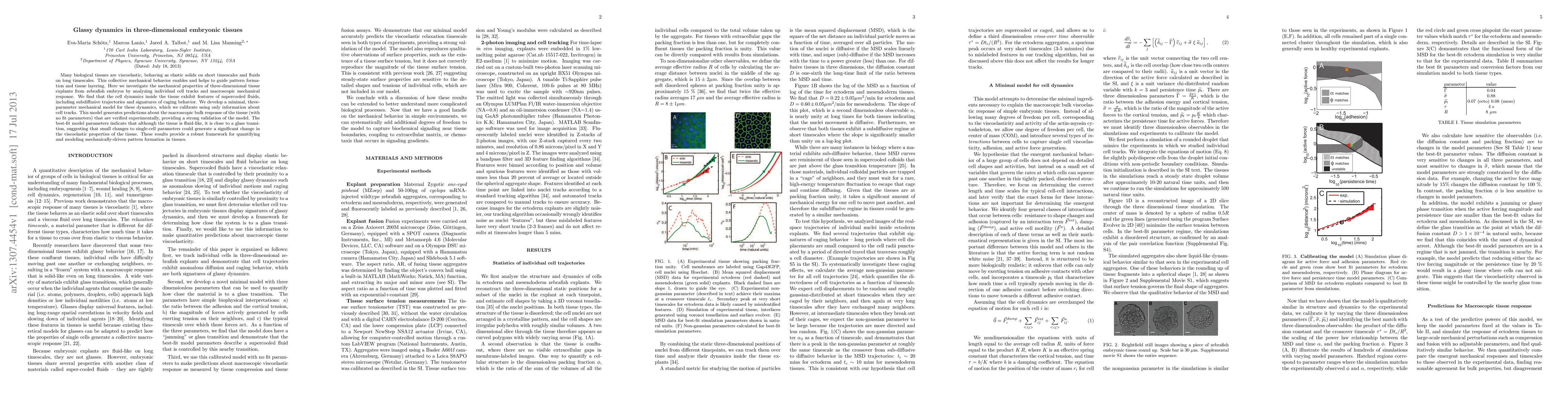
Many biological tissues are viscoelastic, behaving as elastic solids on short timescales and fluids on long timescales. This collective mechanical behavior enables and helps to guide pattern formation and tissue layering. Here we investigate the mechanical properties of three-dimensional tissue explants from zebrafish embryos by analyzing individual cell tracks and macroscopic mechanical response. We find that the cell dynamics inside the tissue exhibit features of supercooled fluids, including subdiffusive trajectories and signatures of caging behavior. We develop a minimal, three-parameter mechanical model for these dynamics, which we calibrate using only information about cell tracks. This model generates predictions about the macroscopic bulk response of the tissue (with no fit parameters) that are verified experimentally, providing a strong validation of the model. The best-fit model parameters indicate that although the tissue is fluid-like, it is close to a glass transition, suggesting that small changes to single-cell parameters could generate a significant change in the viscoelastic properties of the tissue. These results provide a robust framework for quantifying and modeling mechanically-driven pattern formation in tissues.
Seven facets of this paper, analysed and brought into focus by AI.
This research has implications for understanding the behavior of living tissues and their potential applications in biomedicine.
A numerical model was developed to simulate the dynamics of active tissues.
This research has implications for understanding the behavior of living tissues and their potential applications in biomedicine.
A novel numerical method was developed to simulate the dynamics of active tissues, allowing for the prediction of tissue behavior under various conditions.
The use of persistence time as a key parameter in simulating active tissue dynamics is a new approach that has not been explored before.
Current paper (gray), citations (green), references (blue)
Display is limited for performance on very large graphs.
Discussion 0