Graph Neural Network and Superpixel Based Brain Tissue Segmentation (Corrected Version)
Publication
Metrics
AI Quick Summary
This paper proposes a graph neural network (GNN) based method, GNN-SEG, for brain tissue segmentation using superpixels as processing units, enhancing feature integration through interaction modules. Experimental results demonstrate GNN-SEG's superiority over state-of-the-art CNN-based methods on brain MRI datasets.
Paper Preview
Abstract
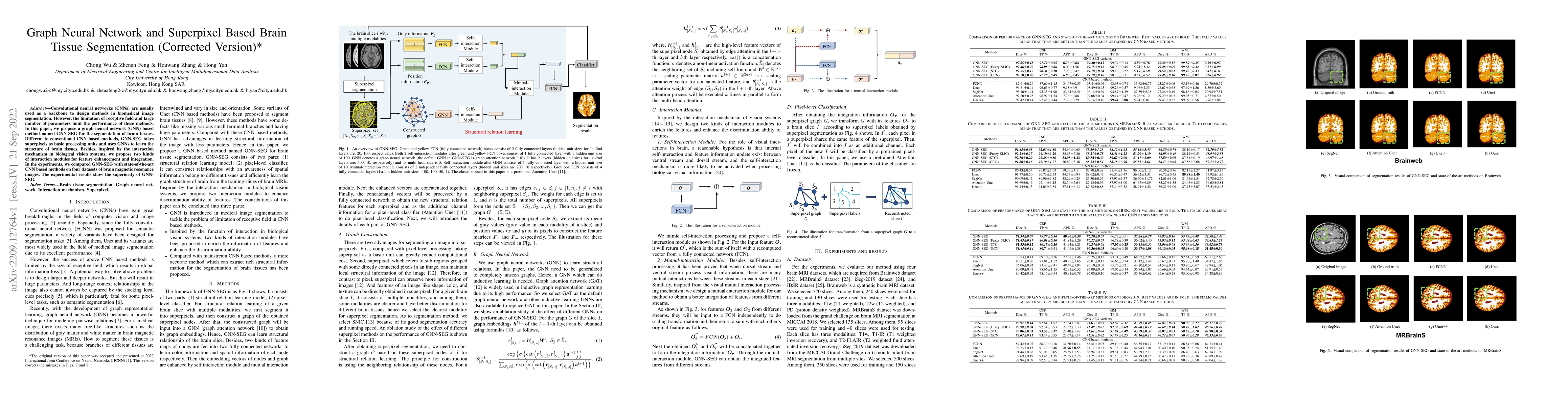
Convolutional neural networks (CNNs) are usually used as a backbone to design methods in biomedical image segmentation. However, the limitation of receptive field and large number of parameters limit the performance of these methods. In this paper, we propose a graph neural network (GNN) based method named GNN-SEG for the segmentation of brain tissues. Different to conventional CNN based methods, GNN-SEG takes superpixels as basic processing units and uses GNNs to learn the structure of brain tissues. Besides, inspired by the interaction mechanism in biological vision systems, we propose two kinds of interaction modules for feature enhancement and integration. In the experiments, we compared GNN-SEG with state-of-the-art CNN based methods on four datasets of brain magnetic resonance images. The experimental results show the superiority of GNN-SEG.
AI Key Findings
Get AI-generated insights about this paper's methodology, results, significance, and more — seven facets brought into focus.
Impact
Paper Details
Authors
PDF Preview
Key Terms
Citation Network
Current paper (gray), citations (green), references (blue)
Display is limited for performance on very large graphs.
Discussion 0