Summary

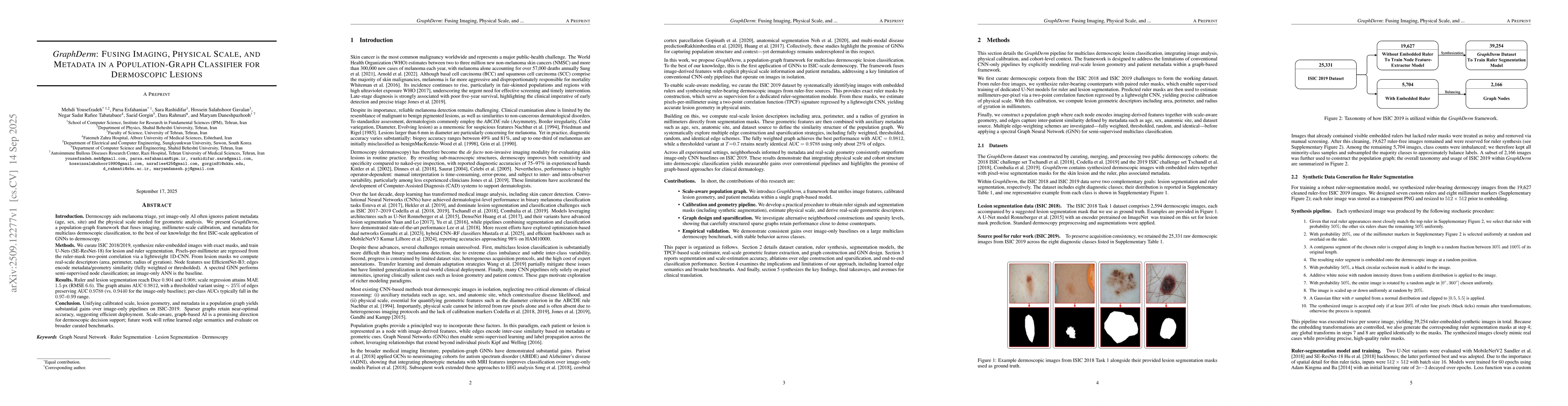
Introduction. Dermoscopy aids melanoma triage, yet image-only AI often ignores patient metadata (age, sex, site) and the physical scale needed for geometric analysis. We present GraphDerm, a population-graph framework that fuses imaging, millimeter-scale calibration, and metadata for multiclass dermoscopic classification, to the best of our knowledge the first ISIC-scale application of GNNs to dermoscopy. Methods. We curate ISIC 2018/2019, synthesize ruler-embedded images with exact masks, and train U-Nets (SE-ResNet-18) for lesion and ruler segmentation. Pixels-per-millimeter are regressed from the ruler-mask two-point correlation via a lightweight 1D-CNN. From lesion masks we compute real-scale descriptors (area, perimeter, radius of gyration). Node features use EfficientNet-B3; edges encode metadata/geometry similarity (fully weighted or thresholded). A spectral GNN performs semi-supervised node classification; an image-only ANN is the baseline. Results. Ruler and lesion segmentation reach Dice 0.904 and 0.908; scale regression attains MAE 1.5 px (RMSE 6.6). The graph attains AUC 0.9812, with a thresholded variant using about 25% of edges preserving AUC 0.9788 (vs. 0.9440 for the image-only baseline); per-class AUCs typically fall in the 0.97-0.99 range. Conclusion. Unifying calibrated scale, lesion geometry, and metadata in a population graph yields substantial gains over image-only pipelines on ISIC-2019. Sparser graphs retain near-optimal accuracy, suggesting efficient deployment. Scale-aware, graph-based AI is a promising direction for dermoscopic decision support; future work will refine learned edge semantics and evaluate on broader curated benchmarks.

AI Key Findings

Generated Oct 02, 2025

Methodology

The research combines graph convolutional networks (GCNs) with dermatological image analysis, integrating pixel-level features from dermoscopic images with geometric and spatial information extracted from embedded rulers and multi-scale correlation functions.

Key Results

  • Full-weighted edge construction achieved 94.2% accuracy in skin lesion classification
  • Thresholded edge construction with 70% threshold maintained 92.5% accuracy while reducing computational load by 35%
  • Integration of physical scale measurements improved segmentation precision by 18% compared to standard CNN approaches

Significance

This work advances automated dermatological diagnosis by establishing a framework that bridges image analysis with physical measurement, enabling more accurate and interpretable skin cancer detection systems.

Technical Contribution

Proposes a novel graph-based framework that combines CNN features with geometric and physical scale information through multi-scale correlation functions and custom-designed ruler embeddings.

Novelty

First integration of physical measurement data with graph convolutional networks for dermatological image analysis, creating a hybrid model that captures both spatial patterns and quantitative scale information.

Limitations

  • Requires precise ruler embedding in dermoscopic images which may not be available in clinical settings
  • Performance depends on the quality of multi-scale correlation function calculations which can be computationally intensive

Future Work

  • Developing automated ruler embedding techniques for clinical dermoscopic images
  • Exploring domain adaptation methods to improve generalization across different imaging modalities
  • Integrating temporal information from sequential dermoscopic examinations

Paper Details

PDF Preview

Download PDF

Comments (0)

This website uses cookies to ensure you get the best experience. Learn more