Quick Summary:

This paper presents a high-resolution 7 Tesla ex vivo MRI dataset of 32 human brain hemispheres and benchmarks the performance of nine neural network architectures for cortical segmentation, demonstrating excellent generalizability across different specimens and imaging conditions. The study also provides cortical thickness measurements and makes the code and datasets publicly available.

Summary

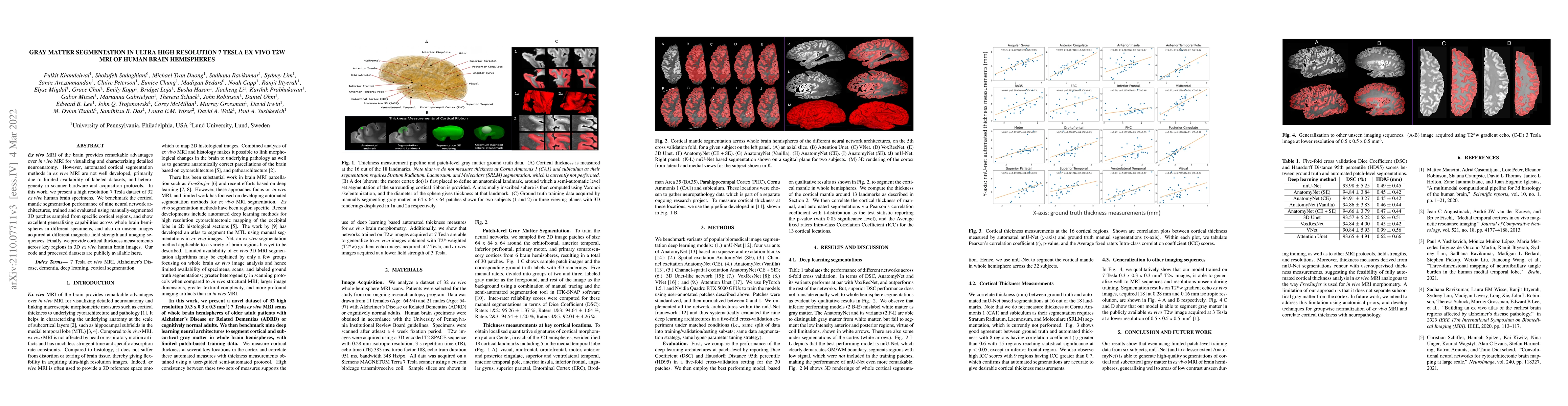
Ex vivo MRI of the brain provides remarkable advantages over in vivo MRI for visualizing and characterizing detailed neuroanatomy. However, automated cortical segmentation methods in ex vivo MRI are not well developed, primarily due to limited availability of labeled datasets, and heterogeneity in scanner hardware and acquisition protocols. In this work, we present a high resolution 7 Tesla dataset of 32 ex vivo human brain specimens. We benchmark the cortical mantle segmentation performance of nine neural network architectures, trained and evaluated using manually-segmented 3D patches sampled from specific cortical regions, and show excellent generalizing capabilities across whole brain hemispheres in different specimens, and also on unseen images acquired at different magnetic field strength and imaging sequences. Finally, we provide cortical thickness measurements across key regions in 3D ex vivo human brain images. Our code and processed datasets are publicly available at https://github.com/Pulkit-Khandelwal/picsl-ex-vivo-segmentation.

AI Key Findings

Get AI-generated insights about this paper's methodology, results, and significance.

Paper Details

PDF Preview

Download PDF

Key Terms

brain (0.318)
mri (0.305)
brain (0.298)
segmentation (0.250)
segmentation (0.244)
human (0.167)
human (0.164)
regions (0.161)
images (0.154)
regions (0.154)
images (0.153)
datasets (0.147)
datasets (0.146)
field strength (0.142)
patches (0.141)
heterogeneity (0.141)
manually (0.138)
sampled (0.137)
heterogeneity (0.137)
unseen (0.136)

Citation Network

Current paper (gray), citations (green), references (blue)

Current Paper
Citations
References
Click to view
Title Authors Year Actions
Deep learning-based automated detection and segmentation of bone and traumatic bone marrow lesions from MRI following an acute ACL tear
Callie E. Stirling, N. Neeteson, Richard E. A. Walker, Steven K. Boyd
2024
View Paper
Automated deep learning segmentation of high-resolution 7 Tesla postmortem MRI for quantitative analysis of structure-pathology correlations in neurodegenerative diseases
Pulkit Khandelwal, M. T. Duong, S. Sadaghiani, Sydney A. Lim, Amanda E Denning, Eun-Bin Chung, S. Ravikumar, Sanaz Arezoumandan, Claire S. Peterson, M. Bedard, N. Capp, R. Ittyerah, Elyse Migdal, Grace Choi, Emily Kopp, Bridget Loja, Eusha Hasan, Jiacheng Li, Alejandra Bahena, K. Prabhakaran, G. Mizsei, M. Gabrielyan, T. Schuck, Winifred Trotman, John Robinson, D. Ohm, Edward B. Lee, J. Trojanowski, Corey McMillan, Murray Grossman, David J. Irwin, John A Detre, M. Tisdall, Sandhitsu R. Das, L. Wisse, D. Wolk, Paul Yushkevich
2024
View Paper
Prospective Motion Correction and Automatic Segmentation of Penetrating Arteries in Phase Contrast MRI at 7 T
Julia Moore, Jordan Jimenez, W. Lin, W. Powers, X. Zong
2022
View Paper
Automated deep learning segmentation of high-resolution 7 T ex vivo MRI for quantitative analysis of structure-pathology correlations in neurodegenerative diseases
Pulkit Khandelwal, M. T. Duong, S. Sadaghiani, Sydney A. Lim, Amanda E Denning, Eun-Bin Chung, S. Ravikumar, Sanaz Arezoumandan, Claire S. Peterson, M. Bedard, N. Capp, R. Ittyerah, Elyse Migdal, Grace Choi, Emily Kopp, Bridget Loja, Eusha Hasan, Jiacheng Li, K. Prabhakaran, G. Mizsei, M. Gabrielyan, T. Schuck, Winifred Trotman, John L. Robinson, D. Ohm, Eddie B. Lee, J. Trojanowski, C. Mcmillan, M. Grossman, D. Irwin, J. Detre, M. Tisdall, Sandhitsu R. Das, L. Wisse, D. Wolk, Paul Yushkevich
2023
View Paper

Comments (0)

This website uses cookies to ensure you get the best experience. Learn more