ArxivLens

Publication

Published: Oct 05, 2025
Categories: cs.AI

Metrics

Source: ArXiv

Quick Answers

What methodology did the authors use?

The research introduces GROK, a multimodal retinal MLLM that jointly processes color fundus photographs (CFP), optical coherence tomography (OCT), and text for clinician-grade diagnosis. It employs a CLIP-style dual-encoder architecture, cross-modal alignment, and LoRA-based knowledge-guided fine-tuning to implement a quantitative-to-qualitative chain-of-thought reasoning framework. More in Methodology →

What are the key results?

GROK surpasses 7B- and 32B-parameter baselines in diagnostic classification, report quality, and fine-grained clinical assessment — It outperforms OpenAI-o3 on key metrics while offering superior readability and clinical explainability compared to models like Lingshu More in Key Results →

Why is this work significant?

This research advances medical diagnostics by enabling more accurate and interpretable analysis of retinal images, which can improve early detection and management of ocular and systemic diseases. The methodology could be extended to other medical imaging modalities. More in Significance →

What are the main limitations?

The study relies on the UK Biobank dataset, which may introduce class imbalance and long-tail disease distribution — CLIP-style pretraining only aligns central-foveal 2D scans with 3D biomarkers, omitting volumetric context More in Limitations →

Paper Preview

Abstract

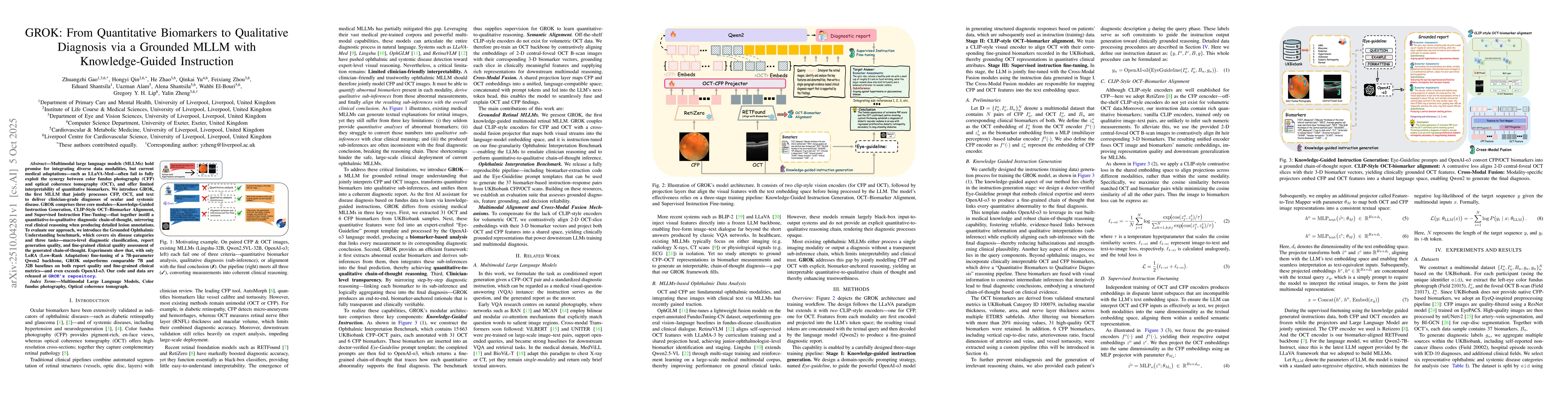
Multimodal large language models (MLLMs) hold promise for integrating diverse data modalities, but current medical adaptations such as LLaVA-Med often fail to fully exploit the synergy between color fundus photography (CFP) and optical coherence tomography (OCT), and offer limited interpretability of quantitative biomarkers. We introduce GROK, a grounded multimodal large language model that jointly processes CFP, OCT, and text to deliver clinician-grade diagnoses of ocular and systemic disease. GROK comprises three core modules: Knowledge-Guided Instruction Generation, CLIP-Style OCT-Biomarker Alignment, and Supervised Instruction Fine-Tuning, which together establish a quantitative-to-qualitative diagnostic chain of thought, mirroring real clinical reasoning when producing detailed lesion annotations. To evaluate our approach, we introduce the Grounded Ophthalmic Understanding benchmark, which covers six disease categories and three tasks: macro-level diagnostic classification, report generation quality, and fine-grained clinical assessment of the generated chain of thought. Experiments show that, with only LoRA (Low-Rank Adaptation) fine-tuning of a 7B-parameter Qwen2 backbone, GROK outperforms comparable 7B and 32B baselines on both report quality and fine-grained clinical metrics, and even exceeds OpenAI o3. Code and data are publicly available in the GROK repository.

Key Findings, in focus

Seven facets of this paper, analysed and brought into focus by AI.

AI-generated · verify against source Generated Oct 11, 2025
In focus · one-line synthesis

This research advances medical diagnostics by enabling more accurate and interpretable analysis of retinal images, which can improve early detection and management of ocular and systemic diseases.

01
MethodologyHow they did it

The research introduces GROK, a multimodal retinal MLLM that jointly processes color fundus photographs (CFP), optical coherence tomography (OCT), and text for clinician-grade diagnosis. It employs a CLIP-style dual-encoder architecture, cross-modal alignment, and LoRA-based knowledge-guided fine-tuning to implement a quantitative-to-qualitative chain-of-thought reasoning framework.

02
ResultsWhat they found
  • GROK surpasses 7B- and 32B-parameter baselines in diagnostic classification, report quality, and fine-grained clinical assessment
  • It outperforms OpenAI-o3 on key metrics while offering superior readability and clinical explainability compared to models like Lingshu
  • The model achieves lower MAE (3.6069 vs 3.8525), lower RMSE (6.1004 vs 6.3583), and higher R2 (0.7106 vs 0.6621) in regression tasks involving OCT biomarkers
03
SignificanceWhy it matters

This research advances medical diagnostics by enabling more accurate and interpretable analysis of retinal images, which can improve early detection and management of ocular and systemic diseases. The methodology could be extended to other medical imaging modalities.

04
Technical ContributionsWhat's new technically

The paper proposes a domain-aligned dual-encoder architecture with cross-modal alignment and knowledge-guided fine-tuning, enabling precise biomarker inference and coherent quantitative-to-qualitative reasoning in retinal analysis.

05
NoveltyWhat sets it apart

This work introduces a CLIP-style dual-encoder framework specifically tailored for retinal analysis, combining OCT and CFP modalities with text inputs through contrastive alignment and LoRA-based fine-tuning for enhanced interpretability and diagnostic accuracy.

06
LimitationsWhat to be cautious about
  • The study relies on the UK Biobank dataset, which may introduce class imbalance and long-tail disease distribution
  • CLIP-style pretraining only aligns central-foveal 2D scans with 3D biomarkers, omitting volumetric context
07
Future WorkWhere it goes next
  • Integrate additional imaging modalities for more comprehensive analysis
  • Explore full-model fine-tuning on larger backbones to further boost performance
  • Conduct extensive clinician evaluations to validate diagnostic accuracy in real-world settings

Paper Details

Paper ID: 2510.04281
Comments: 9 pages, 4 figures, 3 table. Equal contribution: Zhuangzhi Gao and Hongyi Qin. Corresponding author: Yalin Zheng ([email protected])
Categories:
cs.AI

How to Cite This Paper

Reference manager: RIS EndNote


Discussion 0

This website uses cookies to ensure you get the best experience. Learn more