01
MethodologyHow they did it
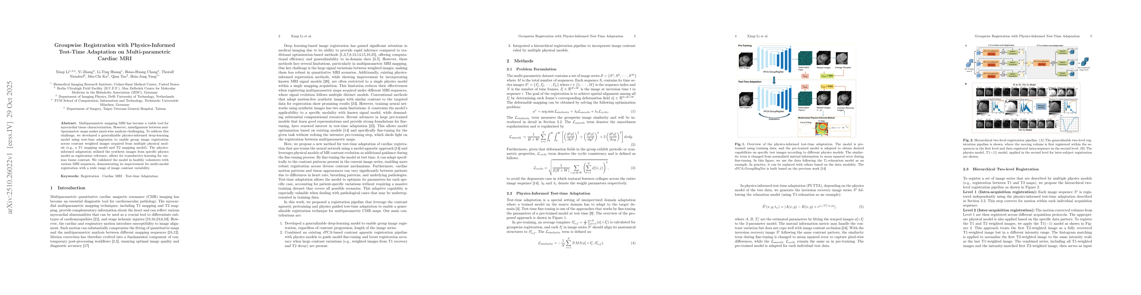
The paper proposes a physics-informed deep learning model with test-time adaptation for groupwise registration of multi-parametric cardiac MRI images. The method uses hierarchical two-level registration, combining intra-acquisition and inter-acquisition registration steps with physics-based models.
Discussion 0