Guiding 3D U-nets with signed distance fields for creating 3D models from images
Publication
Metrics
AI Quick Summary
Researchers propose using signed distance fields to guide deep networks towards creating 3D models from images that resemble real-world anatomy.
Paper Preview
Abstract
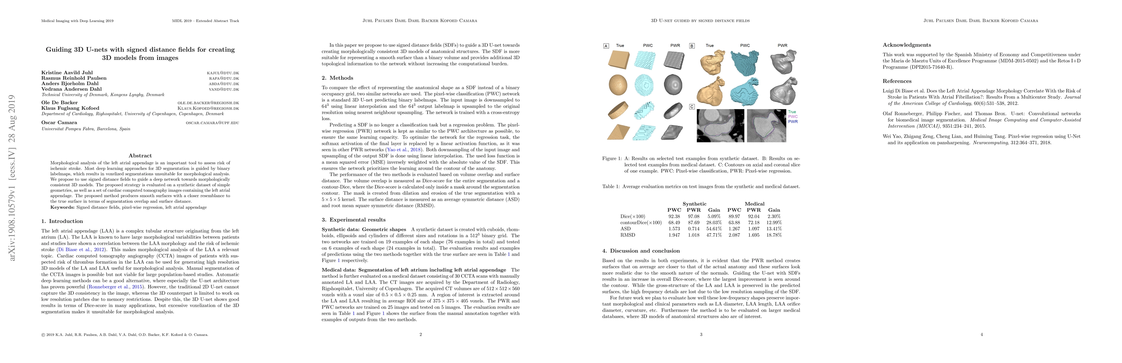
Morphological analysis of the left atrial appendage is an important tool to assess risk of ischemic stroke. Most deep learning approaches for 3D segmentation is guided by binary labelmaps, which results in voxelized segmentations unsuitable for morphological analysis. We propose to use signed distance fields to guide a deep network towards morphologically consistent 3D models. The proposed strategy is evaluated on a synthetic dataset of simple geometries, as well as a set of cardiac computed tomography images containing the left atrial appendage. The proposed method produces smooth surfaces with a closer resemblance to the true surface in terms of segmentation overlap and surface distance.
AI Key Findings
Get AI-generated insights about this paper's methodology, results, significance, and more — seven facets brought into focus.
Impact
Paper Details
Authors
PDF Preview
Key Terms
Citation Network
Current paper (gray), citations (green), references (blue)
Display is limited for performance on very large graphs.
Discussion 0