01
MethodologyHow they did it
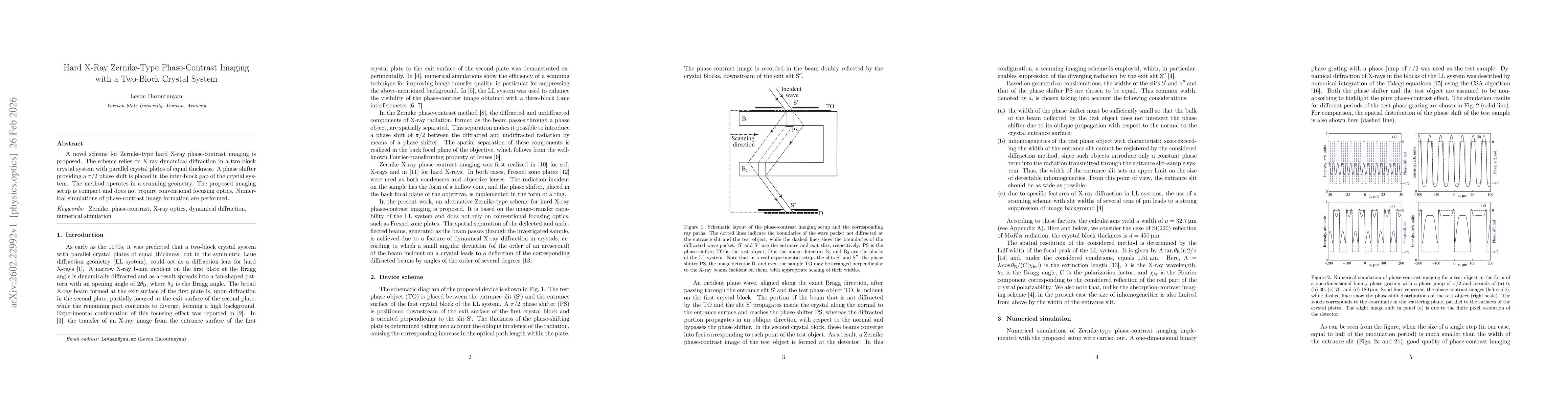
A compact Zernike‑type phase‑contrast imaging scheme was devised using dynamical diffraction in a two‑block Laue‑Laue crystal system with a π/2 phase shifter placed in the inter‑block gap; the setup operates in a scanning geometry without conventional focusing optics and was evaluated through numerical integration of Takagi equations with a CSA algorithm.
Discussion 0