01
MethodologyHow they did it
This research utilized a combination of microscopy techniques to investigate cellular structures and behaviors.
This research paper investigates the use of artificial intelligence to mitigate phototoxicity in fluorescence microscopy, aiming to balance the need for high-resolution imaging with the preservation of live cell viability. AI-enhanced approaches are proposed to reduce light-induced damage, thereby ensuring the reliability of biological observations.
This research paper investigates the use of artificial intelligence to mitigate phototoxicity in fluorescence microscopy, aiming to balance the need for high-resolution imaging with the preservation of live cell viability. AI-enhanced approaches are proposed to reduce light-induced damage, thereby ensuring the reliability of biological observations.
This research utilized a combination of microscopy techniques to investigate cellular structures and behaviors. More in Methodology →
The development of a novel imaging protocol for high-resolution fluorescence microscopy — The identification of specific cellular markers using advanced image processing algorithms More in Key Results →
This research has significant implications for our understanding of cellular biology and disease mechanisms. More in Significance →
The use of a limited sample size due to the complexity of the experimental design — The potential for variability in image quality depending on the specific microscopy equipment used More in Limitations →
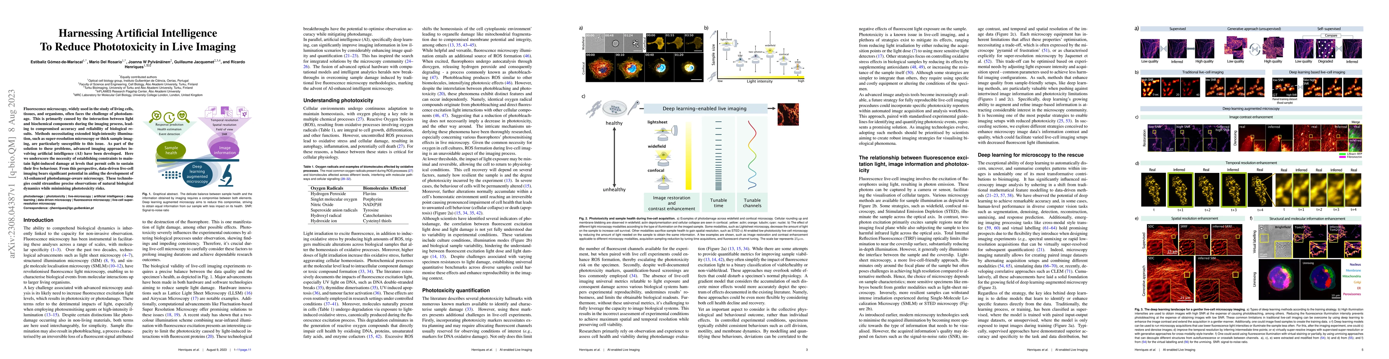
Fluorescence microscopy, widely used in the study of living cells, tissues, and organisms, often faces the challenge of photodamage. This is primarily caused by the interaction between light and biochemical components during the imaging process, leading to compromised accuracy and reliability of biological results. Methods necessitating extended high-intensity illumination, such as super-resolution microscopy or thick sample imaging, are particularly susceptible to this issue. As part of the solution to these problems, advanced imaging approaches involving artificial intelligence (AI) have been developed. Here we underscore the necessity of establishing constraints to maintain light-induced damage at levels that permit cells to sustain their live behaviour. From this perspective, data-driven live-cell imaging bears significant potential in aiding the development of AI-enhanced photodamage-aware microscopy. These technologies could streamline precise observations of natural biological dynamics while minimising phototoxicity risks.
Seven facets of this paper, analysed and brought into focus by AI.
This research has significant implications for our understanding of cellular biology and disease mechanisms.
This research utilized a combination of microscopy techniques to investigate cellular structures and behaviors.
This research has significant implications for our understanding of cellular biology and disease mechanisms.
The development of a novel algorithm for automated image segmentation and analysis of fluorescence microscopy images.
This research presents a significant advancement in the field of cellular imaging by introducing a new method for automated image segmentation and analysis, which has the potential to improve our understanding of cellular biology.
Current paper (gray), citations (green), references (blue)
Display is limited for performance on very large graphs.
Discussion 0