This paper proposes a fully-automated framework for characterizing liver lesions using deep dynamic texture learning on multi-phase CT data, aiming to reduce invasive biopsies. The framework includes a 3D non-isotropic anchor-free detection method and spatially adaptive deep texture learning for precise lesion characterization, achieving an F1 score of 0.763 for differentiating hepatocellular carcinoma from other types, outperforming baseline methods.
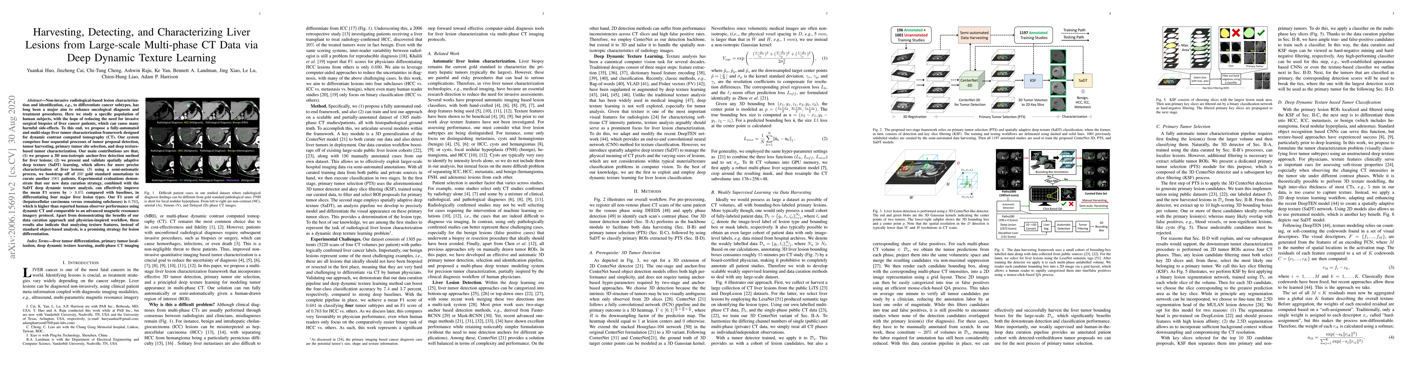
Non-invasive radiological-based lesion characterization and identification, e.g., to differentiate cancer subtypes, has long been a major aim to enhance oncological diagnosis and treatment procedures. Here we study a specific population of human subjects, with the hope of reducing the need for invasive surgical biopsies of liver cancer patients, which can cause many harmful side-effects. To this end, we propose a fully-automated and multi-stage liver tumor characterization framework designed for dynamic contrast computed tomography (CT). Our system comprises four sequential processes of tumor proposal detection, tumor harvesting, primary tumor site selection, and deep texture-based tumor characterization. Our main contributions are that, (1) we propose a 3D non-isotropic anchor-free detection method for liver lesions; (2) we present and validate spatially adaptivedeep texture (SaDT) learning, which allows for more precise characterization of liver lesions; (3) using a semi-automatic process, we bootstrap off of 200 gold standard annotations to curate another 1001 patients. Experimental evaluations demonstrate that our new data curation strategy, combined with the SaDT deep dynamic texture analysis, can effectively improve the mean F1 scores by >8.6% compared with baselines, in differentiating four major liver lesion types. Our F1 score of (hepatocellular carcinoma versus remaining subclasses) is 0.763, which is higher than reported human observer performance using dynamic CT and comparable to an advanced magnetic resonance imagery protocol. Apart from demonstrating the benefits of our data curation approach and physician-inspired workflow, these results also indicate that analyzing texture features, instead of standard object-based analysis, is a promising strategy for lesion differentiation.
Get AI-generated insights about this paper's methodology, results, and significance.
Current paper (gray), citations (green), references (blue)
Lei Zhang, Xu Han, Yuan Gao et al.
Title | Authors | Year | Actions |
---|---|---|---|
Medical Vision Generalist: Unifying Medical Imaging Tasks in Context
|
Sucheng Ren, Xiaoke Huang, Xianhang Li, Junfei Xiao, Jieru Mei, Zeyu Wang, Alan L. Yuille, Yuyin Zhou
|
2024
|
View Paper |
A Cascaded Approach for ultraly High Performance Lesion Detection and False Positive Removal in Liver CT Scans
|
Fakai Wang, Chi-Tung Cheng, Chien-Wei Peng, K. Yan, Min Wu, Le Lu, Chien-Hung Liao, Ling Zhang
|
2023
|
View Paper |
CancerUniT: Towards a Single Unified Model for Effective Detection, Segmentation, and Diagnosis of Eight Major Cancers Using a Large Collection of CT Scans
|
Jieneng Chen, Yingda Xia, Jiawen Yao, K. Yan, Jianpeng Zhang, Le Lu, Fakai Wang, Bo Zhou, Mingyan Qiu, Qihang Yu, Ming Yuan, Wei Fang, Yuxing Tang, Minfeng Xu, Jian Zhou, Yuqian Zhao, Qifeng Wang, X. Ye, Xiaoli Yin, Yu Shi, Xin Chen, Jingren Zhou, A. Yuille, Zai-De Liu, Ling Zhang
|
2023
|
View Paper |
A Knowledge-Guided Framework for Fine-Grained Classification of Liver Lesions Based on Multi-Phase CT Images
|
Xingxin Xu, Qikui Zhu, Hanning Ying, Jiongcheng Li, S. Li, Xiaoqing Liu, Yizhou Yu
|
2022
|
View Paper |
A Unified Multi-Phase CT Synthesis and Classification Framework for Kidney Cancer Diagnosis With Incomplete Data
|
Kwang-Hyun Uhm, Seung‐Won Jung, M. Choi, Sung-Hoo Hong, Sung-Jea Ko
|
2022
|
View Paper |
Context in medical imaging: the case of focal liver lesion classification
|
Moshe Raboh, Dana Levanony, P. Dufort, Arkadiusz Sitek
|
2022
|
View Paper |
Evaluation of an Automated Method to Detect Missed Focal Liver Findings In Single-Phase CT Images of The Abdomen
|
P. Esquinas, Yen-Fu Luo, P. Farzam, Tyler Baldwin, Moshe Raboh, T. Binder, Arkadiusz Sitek, O. Sakhi, Yi-Qing Wang, Sameer Suman, Giovanni Palma, Paul Dufort, Benedikt Graf
|
2022
|
View Paper |
External Attention Assisted Multi-Phase Splenic Vascular Injury Segmentation With Limited Data
|
Yuyin Zhou, D. Dreizin, Yan Wang, Fengze Liu, Wei-lei Shen, A. Yuille
|
2021
|
View Paper |
Hetero-Modal Learning and Expansive Consistency Constraints for Semi-Supervised Detection from Multi-Sequence Data
|
Bolin Lai, Yuhsuan Wu, Xiaoyun Zhou, Peng Wang, Le Lu, Lingyun Huang, Mei Han, Jing Xiao, Heping Hu, Adam P. Harrison
|
2021
|
View Paper |
Fully-Automated Liver Tumor Localization and Characterization from Multi-Phase MR Volumes Using Key-Slice ROI Parsing: A Physician-Inspired Approach
|
Bolin Lai, Xiaoyu Bai, Yuhsuan Wu, Xiaoyun Zhou, Jinzheng Cai, Yuankai Huo, Lingyun Huang, Peng Wang, Yong Xia, Le Lu, Adam P. Harrison, Heping Hu, Jing Xiao
|
2020
|
View Paper |
Towards a Single Unified Model for Effective Detection, Segmentation, and Diagnosis of Eight Major Cancers Using a Large Collection of CT Scans
|
Jieneng Chen, Yingda Xia, Jiawen Yao, Ke Yan, Jianpeng Zhang, Le Lu, Fakai Wang, Bo Zhou, Mingyan Qiu, Qihang Yu, Ming Yuan, Wei Fang, Yuxing Tang, Minfeng Xu, Jian Zhou, Yuqian Zhao, Qifeng Wang, X. Ye, Xiaoli Yin, Yu Shi, Xin Chen, Jingren Zhou, A. L. Yuille, Zai-De Liu, Ling Zhang
|
2023
|
View Paper |
Comments (0)