A non-invasive helmet-based ultrasound brain imager was developed to provide high-resolution brain images in post-hemicraniectomy patients, achieving submillimeter and millisecond spatiotemporal resolutions.
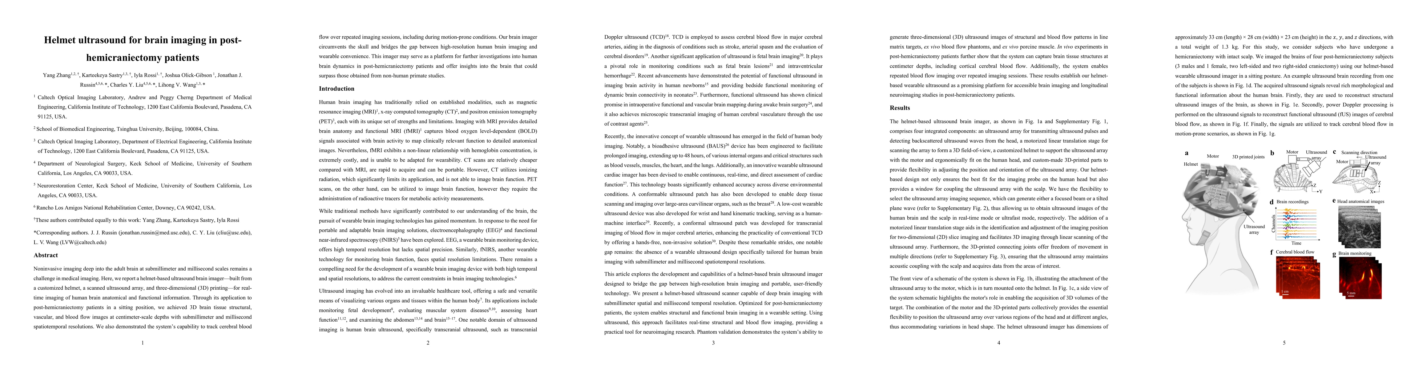
Noninvasive imaging deep into the adult brain at submillimeter and millisecond scales remains a challenge in medical imaging. Here, we report a helmet based ultrasound brain imager built from a customized helmet, a scanned ultrasound array, and three dimensional printing for real time imaging of human brain anatomical and functional information. Through its application to post hemicraniectomy patients in a sitting position, we achieved volumetric brain tissue structural, vascular, and blood flow images at centimeter scale depths with submillimeter and millisecond spatiotemporal resolutions. We also demonstrated the system capability to track cerebral blood flow over repeated imaging sessions, including during motion prone conditions. Our brain imager circumvents the skull and bridges the gap between high resolution human brain imaging and wearable convenience. This imager may serve as a platform for further investigations into human brain dynamics in post hemicraniectomy patients and offer insights into the brain that could surpass those obtained from non human primate studies.
Generated Jun 08, 2025
The research developed a helmet-based ultrasound brain imager using a customized helmet, a scanned ultrasound array, and 3D printing for real-time imaging of human brain anatomical and functional information.
This imager bridges the gap between high-resolution human brain imaging and wearable convenience, potentially offering insights into human brain dynamics surpassing those from non-human primate studies.
The helmet ultrasound brain imager, combining a customized helmet, scanned ultrasound array, and 3D printing, enables real-time, high-resolution imaging of human brain anatomy and function.
This work introduces a wearable, noninvasive ultrasound imaging system for high-resolution brain imaging, addressing a challenge in medical imaging and offering new possibilities for studying human brain dynamics.
Current paper (gray), citations (green), references (blue)
Display is limited for performance on very large graphs.
Michael D. Brown, Christos Strydis, Bastian S. Generowicz et al.
Hamideh Kerdegari, Laura Peralta, Jacqueline Matthew et al.
Waasdorp, R., Munoz-Ibarra, E., Nelissen, F. et al.
No citations found for this paper.
Comments (0)