Authors
Summary
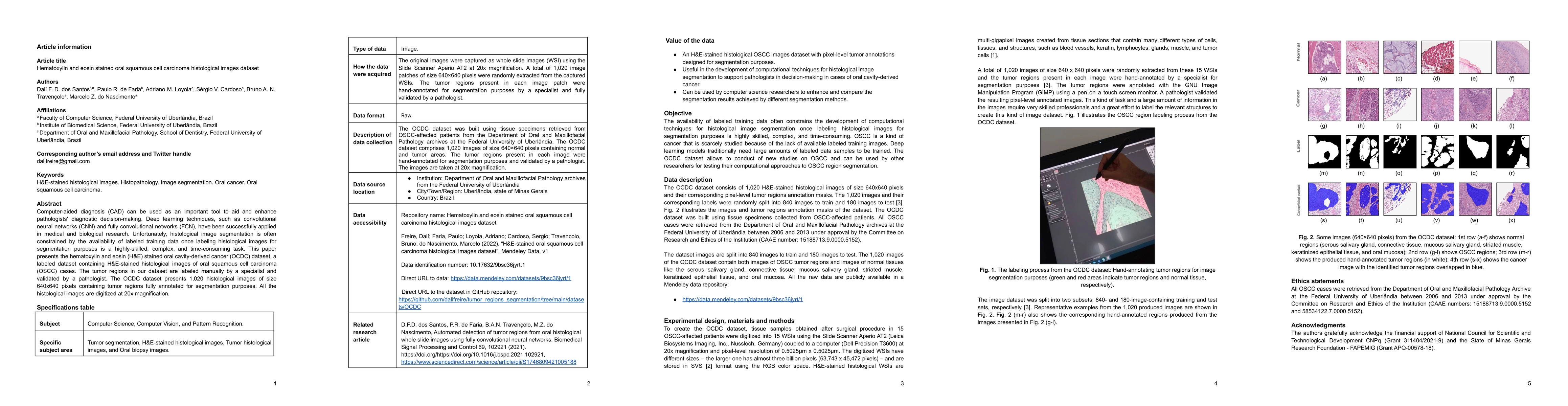
Computer-aided diagnosis (CAD) can be used as an important tool to aid and enhance pathologists' diagnostic decision-making. Deep learning techniques, such as convolutional neural networks (CNN) and fully convolutional networks (FCN), have been successfully applied in medical and biological research. Unfortunately, histological image segmentation is often constrained by the availability of labeled training data once labeling histological images for segmentation purposes is a highly-skilled, complex, and time-consuming task. This paper presents the hematoxylin and eosin (H&E) stained oral cavity-derived cancer (OCDC) dataset, a labeled dataset containing H&E-stained histological images of oral squamous cell carcinoma (OSCC) cases. The tumor regions in our dataset are labeled manually by a specialist and validated by a pathologist. The OCDC dataset presents 1,020 histological images of size 640x640 pixels containing tumor regions fully annotated for segmentation purposes. All the histological images are digitized at 20x magnification.
AI Key Findings
Get AI-generated insights about this paper's methodology, results, and significance.
Paper Details
PDF Preview
Key Terms
Citation Network
Current paper (gray), citations (green), references (blue)
Display is limited for performance on very large graphs.
Similar Papers
Found 4 papersCell type prediction with neighborhood-enhanced cellular embedding using deep learning on hematoxylin and eosin-stained images.
Chen, Yidong, Huang, Yufei, Phan, Nam Nhut et al.
A Cytology Dataset for Early Detection of Oral Squamous Cell Carcinoma
Amit Sethi, Arvind Kumar, Jitender Kumar et al.
Deep Neural Network Pruning for Nuclei Instance Segmentation in Hematoxylin & Eosin-Stained Histological Images
Amirreza Mahbod, Olga Saukh, Rahim Entezari et al.
No citations found for this paper.
Comments (0)