ArxivLens

Quick Summary:

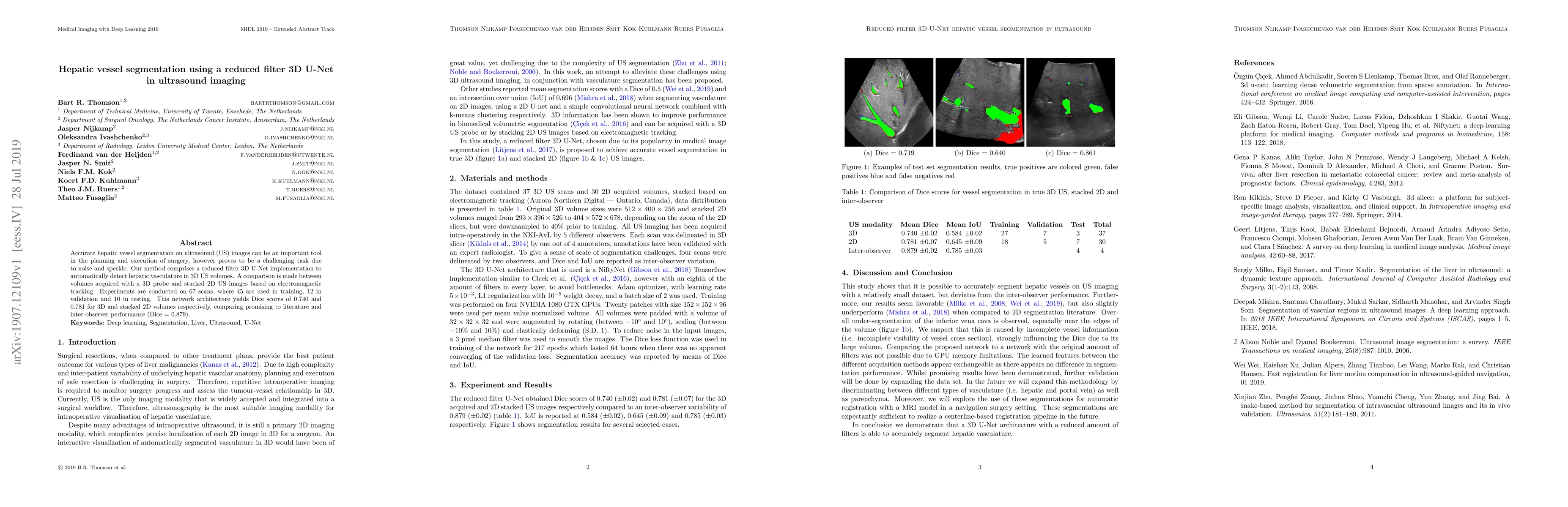
This paper presents a reduced filter 3D U-Net for automatic hepatic vessel segmentation in ultrasound imaging, achieving Dice scores of 0.740 and 0.781 for 3D and 2D volumes respectively, demonstrating competitive performance against inter-observer results.

arXiv Id: 1907.12109
Comments: 3 pages, conference extended abstract. MIDL 2019 [arXiv:1907.08612]
Report No: MIDL/2019/ExtendedAbstract/BklfuXG0KV
Date Published: 2019-07-30
Date Updated: 2019-07-30

Categories:

Key Terms (TF-IDF):

volumes (0.382)
ultrasound (0.202)
images (0.194)
network architecture (0.180)
comprises (0.176)
observer (0.172)
experiments conducted (0.171)
acquired (0.163)
challenging task (0.162)
scores (0.160)
execution (0.158)
planning (0.157)
filter (0.156)
tracking (0.155)
validation (0.153)
net (0.152)
segmentation (0.147)
automatically (0.145)
testing (0.144)
inter (0.143)
License: http://creativecommons.org/licenses/by-sa/4.0/

Summary:

Accurate hepatic vessel segmentation on ultrasound (US) images can be an important tool in the planning and execution of surgery, however proves to be a challenging task due to noise and speckle. Our method comprises a reduced filter 3D U-Net implementation to automatically detect hepatic vasculature in 3D US volumes. A comparison is made between volumes acquired with a 3D probe and stacked 2D US images based on electromagnetic tracking. Experiments are conducted on 67 scans, where 45 are used in training, 12 in validation and 10 in testing. This network architecture yields Dice scores of 0.740 and 0.781 for 3D and stacked 2D volumes respectively, comparing promising to literature and inter-observer performance (Dice = 0.879).

Citation & Reference Network Visualization

This graph shows how this paper is connected to other research. The current paper is shown in dark gray, papers that cite it are in green, and papers it references are in blue. You can:

Current Paper
Papers citing this work (Citations)
Papers cited by this work (References)

• Hover over any node to see the paper title

• Click a node to view that paper's details

• Drag nodes to rearrange the graph

• Use the zoom controls (bottom right) to zoom in/out

Citations and References

Title Authors Year Actions
Progress and clinical translation in hepatocellular carcinoma of deep learning in hepatic vascular segmentation
Tianyang Zhang, Feiyang Yang, Ping Zhang
2024
View Paper
Techniques and Algorithms for Hepatic Vessel Skeletonization in Medical Images: A Survey
Jianfeng Zhang, Fa Wu, Wanru Chang, D. Kong
2022
View Paper
Light-Convolution Dense Selection U-Net (LDS U-Net) for Ultrasound Lateral Bony Feature Segmentation
Sunetra Banerjee, Juan Lyu, Zixun Huang, Ho-fung Leung, T. Lee, De Yang, Steven W. Su, Y. Zheng, S. Ling
2021
View Paper
Sequential U-Net Architecture for Automatic Femoral Artery Segmentation in Ultrasound Images
Ana Estrada Lugo, Niclas Bockelmann, Felix von Haxthausen
2021
View Paper
Segmentation of Liver Anatomy by Combining 3D U-Net Approaches
Abir Affane, Adrian Kucharski, Paul Chapuis, Samuel Freydier, Marie-Ange Lèbre, A. Vacavant, A. Fabijańska
2021
View Paper
Literature Review of Deep Learning Models for Liver Vessels Reconstruction
Abir Affane, Marie-Ange Lèbre, Utkarsh Mittal, A. Vacavant
2020
View Paper
Selecting Image-Guided Surgical Technologies in Oncology: A Surgeon's Perspective.
M. Lindenberg, V. Retèl, J. V. Van Til, K. Kuhlmann, T. Ruers, W. V. van Harten
2020
View Paper
A novel method to model hepatic vascular network using vessel segmentation, thinning, and completion
Xiaoyu Guo, Ruoxiu Xiao, Tao Zhang, Cheng Chen, Jiayu Wang, Zhiliang Wang
2020
View Paper
Medical Image Computing and Computer Assisted Intervention – MICCAI 2020: 23rd International Conference, Lima, Peru, October 4–8, 2020, Proceedings, Part III
Anne L. Martel, P. Abolmaesumi, D. Stoyanov, D. Mateus, Maria A. Zuluaga, S. K. Zhou, Daniel Racoceanu, Leo Joskowicz
2020
View Paper

Comments (0)

This website uses cookies to ensure you get the best experience. Learn more