Publication
Metrics
AI Quick Summary
This paper explores the use of hierarchical Bayesian inference for quantitative assessment of myocardial perfusion MRI, demonstrating improved reliability over traditional least-squares fitting. The Bayesian approach, incorporating prior knowledge and Markov chain Monte Carlo fitting, significantly reduces error and enhances the accuracy of perfusion estimates, as shown through both simulated and patient data.
Paper Preview
Abstract
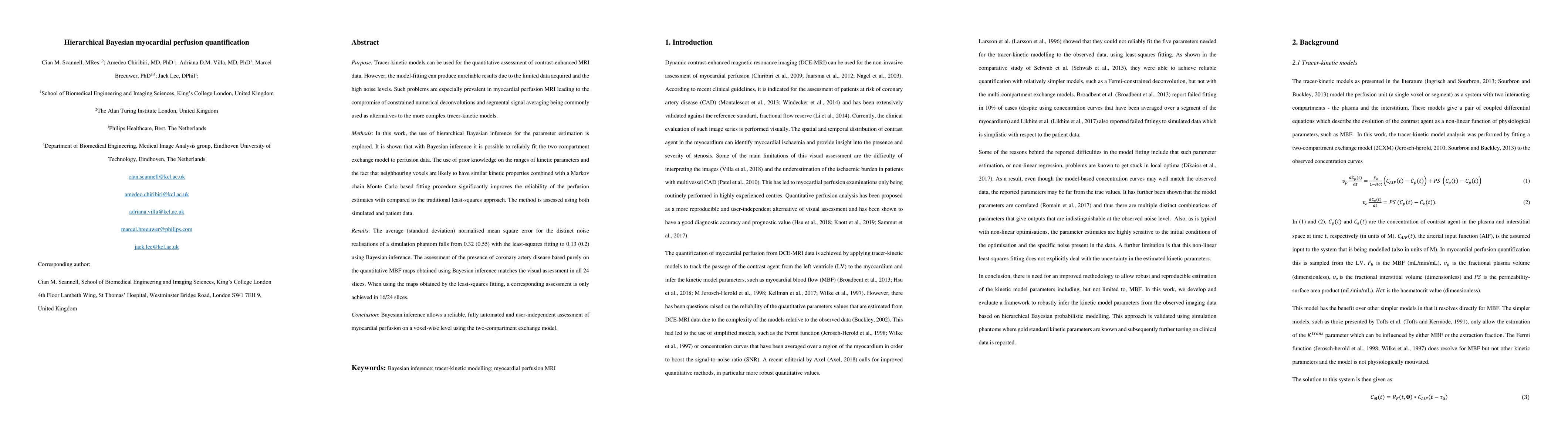
Purpose: Tracer-kinetic models can be used for the quantitative assessment of contrast-enhanced MRI data. However, the model-fitting can produce unreliable results due to the limited data acquired and the high noise levels. Such problems are especially prevalent in myocardial perfusion MRI leading to the compromise of constrained numerical deconvolutions and segmental signal averaging being commonly used as alternatives to the more complex tracer-kinetic models. Methods: In this work, the use of hierarchical Bayesian inference for the parameter estimation is explored. It is shown that with Bayesian inference it is possible to reliably fit the two-compartment exchange model to perfusion data. The use of prior knowledge on the ranges of kinetic parameters and the fact that neighbouring voxels are likely to have similar kinetic properties combined with a Markov chain Monte Carlo based fitting procedure significantly improves the reliability of the perfusion estimates with compared to the traditional least-squares approach. The method is assessed using both simulated and patient data. Results: The average (standard deviation) normalised mean square error for the distinct noise realisations of a simulation phantom falls from 0.32 (0.55) with the least-squares fitting to 0.13 (0.2) using Bayesian inference. The assessment of the presence of coronary artery disease based purely on the quantitative MBF maps obtained using Bayesian inference matches the visual assessment in all 24 slices. When using the maps obtained by the least-squares fitting, a corresponding assessment is only achieved in 16/24 slices. Conclusion: Bayesian inference allows a reliable, fully automated and user-independent assessment of myocardial perfusion on a voxel-wise level using the two-compartment exchange model.
AI Key Findings
Get AI-generated insights about this paper's methodology, results, significance, and more — seven facets brought into focus.
Impact
Paper Details
PDF Preview
Key Terms
Citation Network
Current paper (gray), citations (green), references (blue)
Display is limited for performance on very large graphs.
Discussion 0