The study employs high-contrast interferometric imaging using the evanescent field of a microfiber to detect single-molecule dynamics without labels.
High-Contrast Interferometric Imaging of Single-Molecule Dynamics on Optical Fibers
Publication
Metrics
Quick Answers
What methodology did the authors use?
The study employs high-contrast interferometric imaging using the evanescent field of a microfiber to detect single-molecule dynamics without labels. More in Methodology →
What are the key results?
38 dB SNR enhancement over conventional fluorescence methods — Real-time monitoring of conformational transitions and binding dynamics of proteins More in Key Results →
Why is this work significant?
This approach enables ultra-sensitive, label-free single-molecule analysis with potential applications in biosensing and fundamental biological research. More in Significance →
What are the main limitations?
Temporal resolution limited by camera frame rate — Requires specialized equipment for hyperspectral imaging More in Limitations →
Abstract
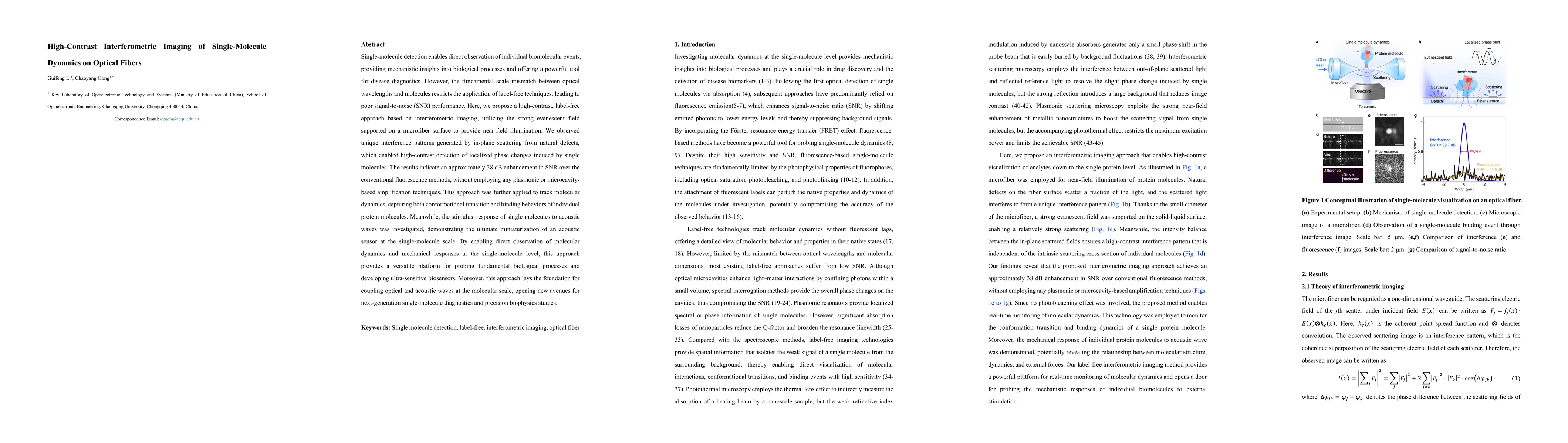
Single-molecule detection enables direct observation of individual biomolecular events, providing mechanistic insights into biological processes and offering a powerful tool for disease diagnostics. However, the fundamental scale mismatch between optical wavelengths and molecules restricts the application of label-free techniques, leading to poor signal-to-noise (SNR) performance. Here, we propose a high-contrast, label-free approach based on interferometric imaging, utilizing the strong evanescent field supported on a microfiber surface to provide near-field illumination. We observed unique interference patterns generated by in-plane scattering from natural defects, which enabled high-contrast detection of localized phase changes induced by single molecules. The results indicate an approximately 38 dB enhancement in SNR over the conventional fluorescence methods, without employing any plasmonic or microcavity-based amplification techniques. This approach was further applied to track molecular dynamics, capturing both conformational transition and binding behaviors of individual protein molecules. Meanwhile, the stimulus-response of single molecules to acoustic waves was investigated, demonstrating the ultimate miniaturization of an acoustic sensor at the single-molecule scale. By enabling direct observation of molecular dynamics and mechanical responses at the single-molecule level, this approach provides a versatile platform for probing fundamental biological processes and developing ultra-sensitive biosensors. Moreover, this approach lays the foundation for coupling optical and acoustic waves at the molecular scale, opening new avenues for next-generation single-molecule diagnostics and precision biophysics studies.
Key Findings, in focus
Seven facets of this paper, analysed and brought into focus by AI.
This approach enables ultra-sensitive, label-free single-molecule analysis with potential applications in biosensing and fundamental biological research.
- 38 dB SNR enhancement over conventional fluorescence methods
- Real-time monitoring of conformational transitions and binding dynamics of proteins
- Demonstration of single-molecule acoustic sensing with frequency response matching theoretical predictions
This approach enables ultra-sensitive, label-free single-molecule analysis with potential applications in biosensing and fundamental biological research.
Creation of a label-free interferometric imaging platform with enhanced signal-to-noise ratio for single-molecule studies
First demonstration of acoustic stimulation-response at single-molecule scale using interferometric imaging without plasmonic or microcavity amplification
- Temporal resolution limited by camera frame rate
- Requires specialized equipment for hyperspectral imaging
- Development of higher frame rate imaging systems
- Integration with other molecular detection techniques
Discussion 0