High-Fidelity 3D Lung CT Synthesis in ARDS Swine Models Using Score-Based 3D Residual Diffusion Models
Publication
Metrics
AI Quick Summary
This study employs a score-based 3D residual diffusion model to synthesize high-fidelity 3D lung CT images from 2D X-ray images and physiological parameters in ARDS swine models, aiming to improve ARDS management without the need for conventional CT scans. The preliminary results show promising accuracy in the synthesized 3D CT images, potentially offering a safer alternative for detailed lung pathology assessment.
Paper Preview
Abstract
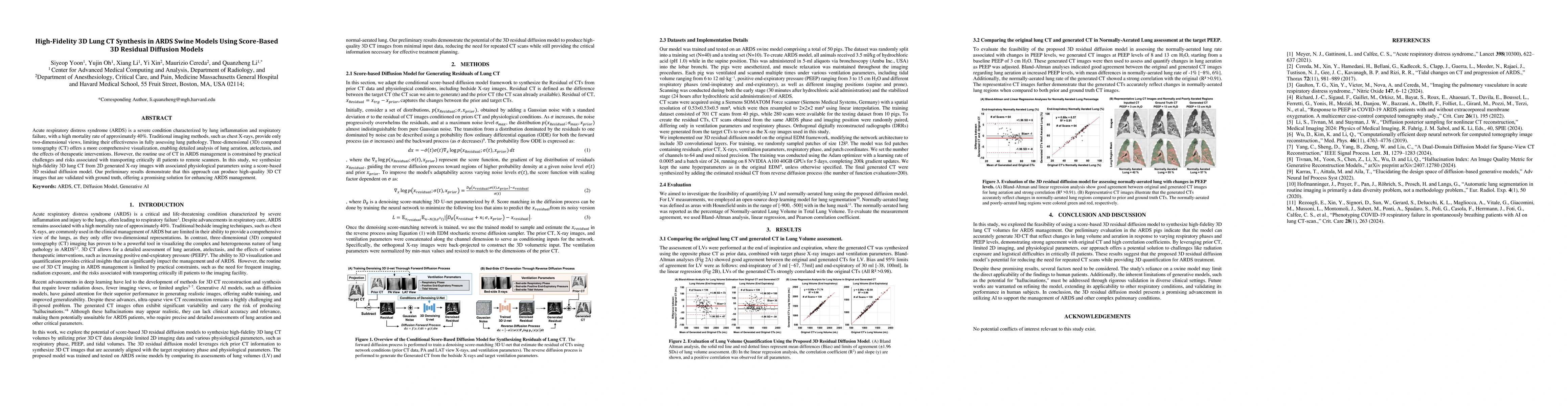
Acute respiratory distress syndrome (ARDS) is a severe condition characterized by lung inflammation and respiratory failure, with a high mortality rate of approximately 40%. Traditional imaging methods, such as chest X-rays, provide only two-dimensional views, limiting their effectiveness in fully assessing lung pathology. Three-dimensional (3D) computed tomography (CT) offers a more comprehensive visualization, enabling detailed analysis of lung aeration, atelectasis, and the effects of therapeutic interventions. However, the routine use of CT in ARDS management is constrained by practical challenges and risks associated with transporting critically ill patients to remote scanners. In this study, we synthesize high-fidelity 3D lung CT from 2D generated X-ray images with associated physiological parameters using a score-based 3D residual diffusion model. Our preliminary results demonstrate that this approach can produce high-quality 3D CT images that are validated with ground truth, offering a promising solution for enhancing ARDS management.
AI Key Findings
Get AI-generated insights about this paper's methodology, results, significance, and more — seven facets brought into focus.
Impact
Paper Details
Authors
PDF Preview
Citation Network
Current paper (gray), citations (green), references (blue)
Display is limited for performance on very large graphs.
Discussion 0