This research introduces high-resolution bi-directional x-ray phase contrast imaging using two-dimensional Talbot array illuminators (TAIs), achieving micrometer-scale resolution and demonstrating potential for non-destructive, three-dimensional imaging of soft tissues, including virtual histology. The TAIs offer superior modulation and flexibility for various x-ray applications.
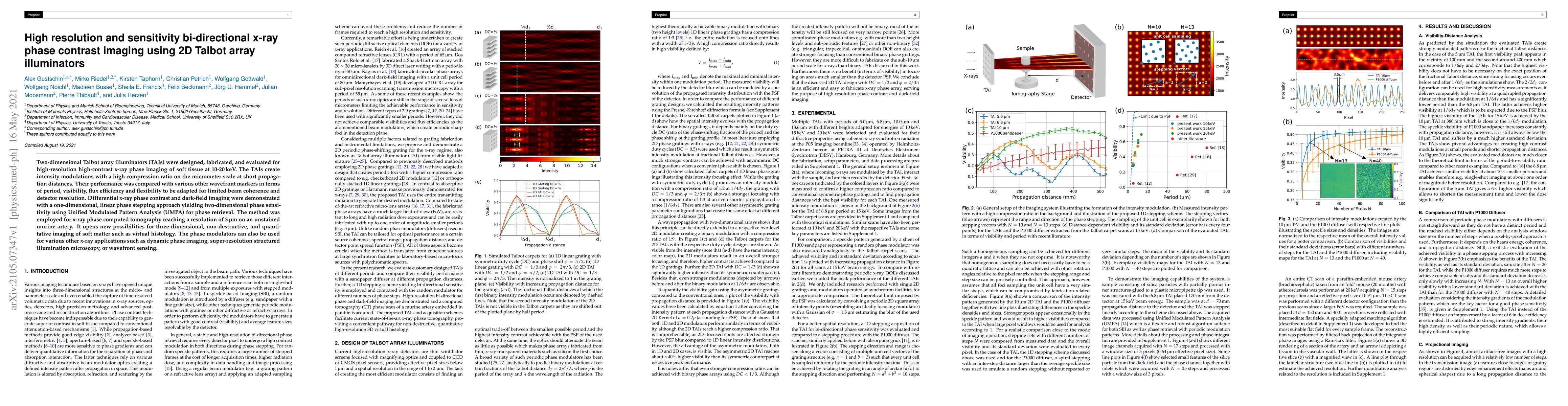
Two-dimensional Talbot array illuminators (TAIs) were designed, fabricated, and evaluated for high-resolution high-contrast x-ray phase imaging of soft tissue at 10-20keV. The TAIs create intensity modulations with a high compression ratio on the micrometer scale at short propagation distances. Their performance was compared with various other wavefront markers in terms of period, visibility, flux efficiency and flexibility to be adapted for limited beam coherence and detector resolution. Differential x-ray phase contrast and dark-field imaging were demonstrated with a one-dimensional, linear phase stepping approach yielding two-dimensional phase sensitivity using Unified Modulated Pattern Analysis (UMPA) for phase retrieval. The method was employed for x-ray phase computed tomography reaching a resolution of 3$\mu$m on an unstained murine artery. It opens new possibilities for three-dimensional, non-destructive, and quantitative imaging of soft matter such as virtual histology. The phase modulators can also be used for various other x-ray applications such as dynamic phase imaging, super-resolution structured illumination microscopy, or wavefront sensing.
Get AI-generated insights about this paper's methodology, results, and significance.
Current paper (gray), citations (green), references (blue)
Michela Esposito, Alessandro Olivo, Marco Endrizzi et al.
David Bate, Alessandro Olivo, Marco Endrizzi et al.
Title | Authors | Year | Actions |
---|---|---|---|
High-resolution X-ray phase-contrast tomography of human placenta with different wavefront markers
|
Sara Savatović, Davis J. Laundon, F. De Marco, Mirko Riedel, J. U. Hammel, M. Busse, Murielle Salomé, Lorella Pascolo, Irene Zanette, Rohan M. Lewis, Julia Herzen, Pierre Thibault
|
2025
|
View Paper |
Centimeter-sized Objects at Micrometer Resolution: Extending Field-of-View in Wavefront Marker X-ray Phase-Contrast Tomography
|
Dominik John, Junan Chen, Christoph Gassner, Sara Savatovi'c, L. Petzold, Sami Wirtensohn, Mirko Riedel, Jörg U. Hammel, J. Moosmann, Felix Beckmann, Matthias Wieczorek, Julia Herzen
|
2024
|
View Paper |
Micro-reinforced Polymer Composite materials studied by Correlative X-ray Imaging
|
A. Mikhaylov, Jorge Luis Beltran Diaz, M. Zakharova, V. Vlnieska, Daniel Münch, Edwin Fohtung, S. Pezzin, D. Kunka
|
2024
|
View Paper |
Helical sample-stepping for faster speckle-based multi-modal tomography with the Unified Modulated Pattern Analysis (UMPA) model
|
Sara Savatović, F. De Marco, Mirko Riedel, V. Di Trapani, Marco Margini, Ginevra Lautizi, Jörg U. Hammel, Julia Herzen, Pierre Thibault
|
2023
|
View Paper |
An Ultra-Fast Temporal Talbot Array Illuminator
|
Manuel P. Fernández, Saket Kaushal, B. Crockett, L. A. Bulus-Rossini, P. Costanzo‐Caso, J. Azaña
|
2023
|
View Paper |
Comparing x-ray phase-contrast imaging using a Talbot array illuminator to propagation-based imaging for non-homogeneous biomedical samples
|
M. Riedel, Kirsten Taphorn, A. Gustschin, M. Busse, J. Hammel, J. Moosmann, F. Beckmann, F. Fischer, P. Thibault, J. Herzen
|
2023
|
View Paper |
Cardiopulmonary structural, functional and immune-alterations in a Down syndrome mouse model and upon modulation of EGCG
|
B. Tielemans, Sergi Llambrich, Laura Seldeslachts, J. Cremer, Hung-Chang Tsui, Anne-Charlotte Jonckheere, Fopke Marain, M. Riedel, Jens Wouters, J. Herzen, B. Leszczyński, E. Verbeken, J. Vanoirbeek, G. Velde
|
2023
|
View Paper |
On the quantification of sample microstructure using single-exposure x-ray dark-field imaging via a single-grid setup
|
Y. Y. How, D. Paganin, K. Morgan
|
2022
|
View Paper |
Three-dimensional analyses of vascular network morphology in a murine lymph node by X-ray phase-contrast tomography with a 2D Talbot array
|
F. Schwarzenberg, Paula Schütz, J. Hammel, M. Riedel, Jasmin Bartl, S. Bordbari, Svea-Celina Frank, Bernd Walkenfort, M. Busse, J. Herzen, C. Lohr, Clemens Wülfing, S. Henne
|
2022
|
View Paper |
Directional dark-field retrieval with single-grid x-ray imaging.
|
M. Croughan, Y. Y. How, Allan Pennings, K. Morgan
|
2022
|
View Paper |
High-speed processing of X-ray wavefront marking data with the Unified Modulated Pattern Analysis (UMPA) model.
|
F. De Marco, Sara Savatović, Ronan Smith, V. Di Trapani, Marco Margini, Ginevra Lautizi, P. Thibault
|
2022
|
View Paper |
Inverted Hartmann mask made by deep X-ray lithography for single-shot multi-contrast X-ray imaging with laboratory setup.
|
A. Mikhaylov, M. Zakharova, V. Vlnieska, Ankita Khanda, Sabine Bremer, M. Zuber, Sergio Henrique Pezzin, D. Kunka
|
2022
|
View Paper |
Quantifying the x-ray dark-field signal in single-grid imaging.
|
Y. Y. How, K. Morgan
|
2021
|
View Paper |
Comments (0)