Quick Answers
What methodology did the authors use?
The study employs a multimode optical fiber-based endoscopy system combined with fluorescence lifetime imaging (FLIm) and machine learning algorithms to analyze tissue autofluorescence for cancer detection. More in Methodology →
What are the key results?
Successful differentiation between precancerous, cancerous, and healthy oral tissues using multispectral autofluorescence lifetime imaging endoscopy — Development of a machine learning model achieving high accuracy in tissue classification More in Key Results →
Why is this work significant?
This research advances minimally invasive cancer diagnostics by enabling real-time, high-resolution tissue analysis during surgery, potentially improving patient outcomes through more precise tumor removal. More in Significance →
What are the main limitations?
Limited to oral tissues and may require adaptation for other cancer types — Dependence on high-quality fluorescence signals which may be affected by tissue heterogeneity More in Limitations →
Abstract
Endoscopic optical imaging using a single multimode fiber (MMF) has emerged as a promising approach for highly compact, minimally invasive, and high-resolution imaging. Unlike conventional fiber bundles, MMF-based endomicroscopes exploit the controlled excitation of multiple spatially overlapping modes in a single MMF. of core diameters of tens of micrometers. to deliver and collect light to form images with sub-micrometer resolution. Here, we introduce a fluorescence lifetime imaging microscopy (FLIM) modality to the MMF endomicroscope. We use amplitude modulation of a 405 nm single-mode light source at radio frequency (RF) and lock-in detection of autofluorescence to obtain intensity and lifetime images at sub-micrometer resolution. We experimentally demonstrate the capability of the ultrathin endomicroscope to perform label-free imaging in thick ex vivo murine submandibular gland tissue. With a temporal resolution of 0.03 ns, the FLIM images show distinguished structures of lifetime differences down to 0.5 ns. The combination of sub-micrometer fluorescence intensity and lifetime images in a minimally invasive endomicroscope opens new avenues for label-free cancer detection.
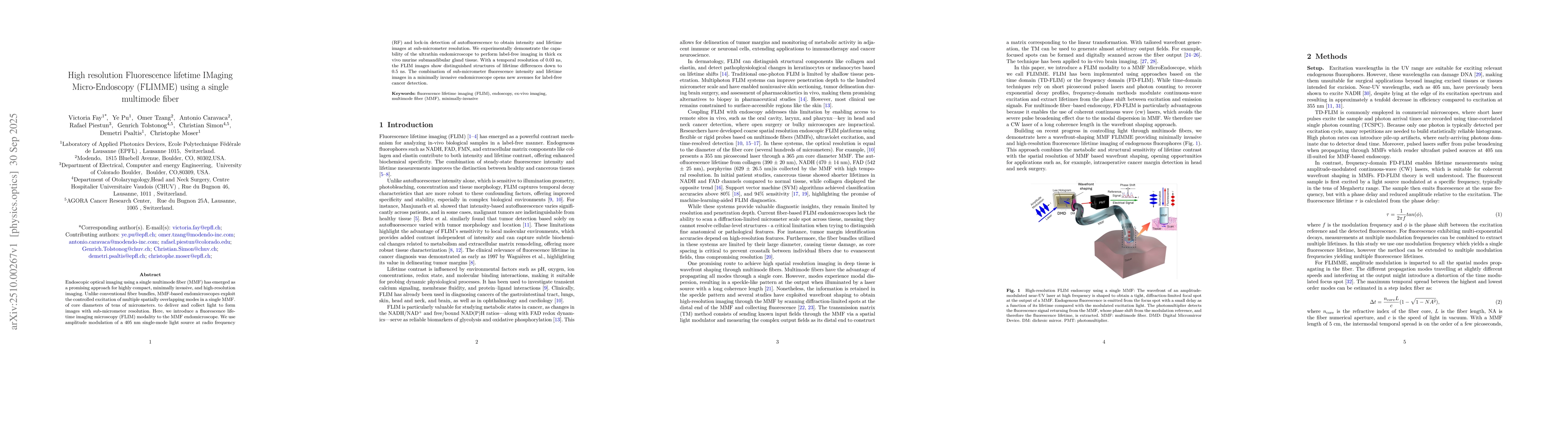
AI Key Findings
Generated Oct 05, 2025
Methodology — What approach did the authors take?
The study employs a multimode optical fiber-based endoscopy system combined with fluorescence lifetime imaging (FLIm) and machine learning algorithms to analyze tissue autofluorescence for cancer detection.
Key Results — What are the main findings?
- Successful differentiation between precancerous, cancerous, and healthy oral tissues using multispectral autofluorescence lifetime imaging endoscopy
- Development of a machine learning model achieving high accuracy in tissue classification
- Demonstration of the potential for real-time intraoperative guidance in head and neck cancer surgery
Significance — Why does this research matter?
This research advances minimally invasive cancer diagnostics by enabling real-time, high-resolution tissue analysis during surgery, potentially improving patient outcomes through more precise tumor removal.
Technical Contribution — What is the technical contribution?
Introduction of a lensless, high-resolution endoscopic system using digital scanning through multimode fibers for fluorescence lifetime imaging
Novelty — What is new about this work?
Combines multimode fiber endoscopy with machine learning for real-time cancer detection, offering a compact, non-invasive solution for intraoperative tissue analysis
Limitations — What are the limitations of this study?
- Limited to oral tissues and may require adaptation for other cancer types
- Dependence on high-quality fluorescence signals which may be affected by tissue heterogeneity
Future Work — What did the authors propose for future work?
- Validation in clinical settings with diverse patient populations
- Integration with other imaging modalities for multimodal diagnosis
- Development of portable systems for broader clinical adoption
How to Cite This Paper
@article{caravaca2025high,
title = {High resolution Fluorescence lifetime IMaging Micro-Endoscopy (FLIMME)
using a single multimode fiber},
author = {Caravaca, Antonio and Simon, Christian and Moser, Christophe and others},
year = {2025},
eprint = {2510.00267},
archivePrefix = {arXiv},
primaryClass = {physics.optics},
}
Caravaca, A., Simon, C., Moser, C., Psaltis, D., Tolstonog, G., Tzang, O., Piestun, R., Fay, V., & Pu, Y. (2025). High resolution Fluorescence lifetime IMaging Micro-Endoscopy (FLIMME)
using a single multimode fiber. arXiv. https://arxiv.org/abs/2510.00267
Caravaca, Antonio, et al. "High resolution Fluorescence lifetime IMaging Micro-Endoscopy (FLIMME)
using a single multimode fiber." arXiv, 2025, arxiv.org/abs/2510.00267.
PDF Preview
Similar Papers
Found 4 papersHigh-Resolution Imaging of Cold Atoms through a Multimode Fiber
Kilian Müller, Nicolas Vitrant, Sébastien Garcia et al.
Flexible single multimode fiber imaging using white LED
Minyu Fan, Yu Cao, Sha Wang et al.
Toward Micro-Endoscopy: Distal-Free, Configuration-Agnostic Focusing Through Multimode Fiber
Dvir Marsh, Lior Fridman, Stav Lotan et al.
Super-resolution Live-cell Fluorescence Lifetime Imaging
Raphaël Marchand, Henning Ortkrass, Daniel Aziz et al.
Comments (0)