ArxivLens

Quick Summary:

This paper introduces high-resolution hard X-ray magnetic imaging using dichroic ptychography, achieving 45 nm resolution to visualize the magnetic domain configuration in a FeGd multilayer. This technique enables detailed 3D analysis of micrometer-sized systems, paving the way for nanoscale investigations of buried magnetic structures.

Journal Reference: Phys. Rev. B 94, 064421 (2016)
DOI: 10.1103/PhysRevB.94.064421
arXiv Id: 1603.03588
Date Published: 2016-08-24
Date Updated: 2016-08-24

Categories:

Key Terms (TF-IDF):

imaging (0.299)
magnetic (0.293)
nanoscale (0.282)
ray (0.232)
resolution (0.228)
hard (0.217)
edge (0.208)
spatial (0.187)
reaching (0.169)
magnetic properties (0.168)
sized (0.162)
spanning (0.158)
advance (0.157)
circular (0.156)
spectroscopic (0.146)
nm (0.144)
fe (0.140)
systems (0.136)
investigation (0.133)
influence (0.132)
License: http://arxiv.org/licenses/nonexclusive-distrib/1.0/

Summary:

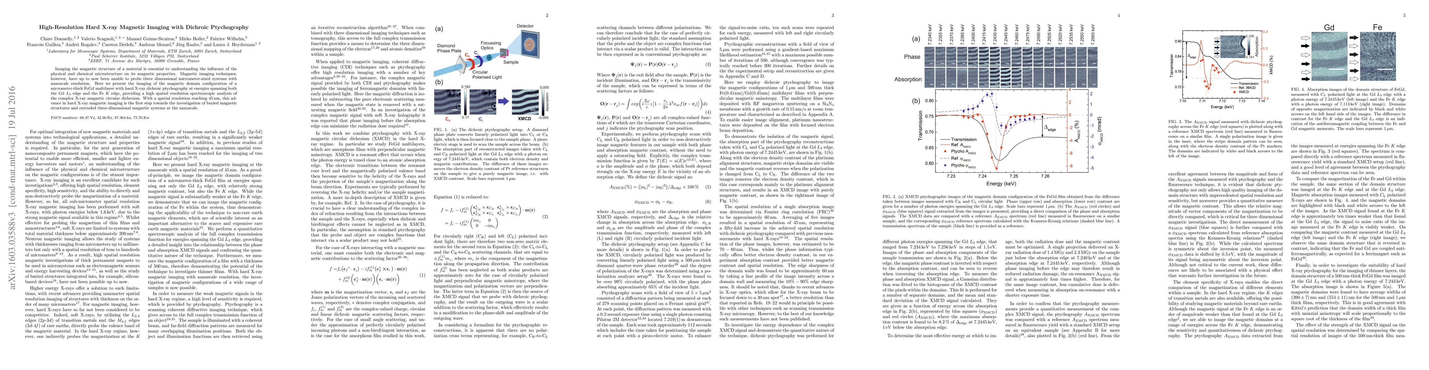
Imaging the magnetic structure of a material is essential to understanding the influence of the physical and chemical microstructure on its magnetic properties. Magnetic imaging techniques, however, have up to now been unable to probe 3D micrometer-sized systems with nanoscale resolution. Here we present the imaging of the magnetic domain configuration of a micrometre-thick FeGd multilayer with hard X-ray dichroic ptychography at energies spanning both the Gd L3 edge and the Fe K edge, providing a high spatial resolution spectroscopic analysis of the complex X-ray magnetic circular dichroism. With a spatial resolution reaching 45 nm, this advance in hard X-ray magnetic imaging is the first step towards the investigation of buried magnetic structures and extended three-dimensional magnetic systems at the nanoscale.

Citation & Reference Network Visualization

This graph shows how this paper is connected to other research. The current paper is shown in dark gray, papers that cite it are in green, and papers it references are in blue. You can:

Current Paper
Papers citing this work (Citations)
Papers cited by this work (References)

• Hover over any node to see the paper title

• Click a node to view that paper's details

• Drag nodes to rearrange the graph

• Use the zoom controls (bottom right) to zoom in/out

Citations and References

Title Authors Year Actions
Electronvolt energy resolution with broadband ptychography
Silvia Cipiccia, Wiebe Stolp, Luca Fardin, R. Ziesche, Ingo Manke, Matthieu Boone, Chris Armstrong, Joachim R. Binder, N. Bohn, Alessandro Olivo, D. Batey
2024
View Paper
Magnetic X-Ray Imaging Using a Single Polarization and Multimodal Ptychography.
M. Di Pietro Martínez, A. Wartelle, Nicolas Mille, Stefan Stanescu, Rachid Belkhou, Farid Fettar, Vincent Favre-Nicolin, Guillaume Beutier
2024
View Paper
Small-angle X-ray resonant magnetic scattering at the Co M$_{2,3}$ and L$_3$ edges observed with photoemission electron microscopy
A. Wartelle, Marisel Di Pietro Mart'inez, O. Fruchart, Philippe David, Guillaume Beutier
2024
View Paper
Soft X-Ray Phase Nanomicroscopy of Micrometer-Thick Magnets
Jeffrey Neethirajan, B. Daurer, Marisel Di Pietro Mart'inez, A. Hrabec, Luke Turnbull, R. Yamamoto, Marina Raboni Ferreira, A. Štefančič, D. Mayoh, G. Balakrishnan, Zhaowen Pei, Pengfei Xue, Liao Chang, Emilie Ringe, Richard J. Harrison, S. Valencia, Majid Kazemian, B. Kaulich, C. Donnelly
2023
View Paper
Micromagnetics of magnetic chemical modulations in soft-magnetic cylindrical nanowires
L. Álvaro-Gómez, S. Ruiz-G'omez, C. Fern'andez-Gonz'alez, M. Schöbitz, N. Mille, J. Hurst, D. Tiwari, A. De Riz, I. M. Andersen, J. Bachmann, L. Cagnon, M. Foerster, L. Aballe, R. Belkhou, J. Toussaint, C. Thirion, A. Masseboeuf, D. Gusakova, L. P'erez, O. Fruchart
2022
View Paper
X-ray Spectroptychography
S. Urquhart
2022
View Paper
Maskless Fourier transform holography.
K. Keskinbora, A. Levitan, R. Comin
2021
View Paper
X-ray Microspectroscopy and Ptychography on Nanoscale Structures in Rock Varnish
Jan‐David Förster, I. Bykova, Dorothea S. Macholdt, K. Jochum, M. Kappl, A. Kilcoyne, Maren Müller, Antje Sorowka, B. Weber, M. Weigand, G. Schütz, M. Andreae, C. Pöhlker
2021
View Paper
Aberration characterization of x-ray optics using multi-modal ptychography and a partially coherent source
T. Moxham, D. Laundy, V. Dhamgaye, O. Fox, K. Sawhney, A. Korsunsky
2021
View Paper
Maskless off-axis X-ray Holography
K. Keskinbora, A. Levitan, G. Schutz, R. Comin
2021
View Paper
Ptychographic imaging and micromagnetic modeling of thermal melting of nanoscale magnetic domains in antidot lattices
J. Gräfe, M. Skripnik, G. Dieterle, F. Haering, M. Weigand, I. Bykova, N. Träger, H. Stoll, T. Tyliszczak, D. Vine, P. Ziemann, U. Wiedwald, D. Shapiro, U. Nowak, G. Schütz, E. Goering
2020
View Paper
An ultrahigh-resolution soft x-ray microscope for quantitative analysis of chemically heterogeneous nanomaterials
D. Shapiro, S. Babin, R. Celestre, W. Chao, R. Conley, P. Denes, B. Enders, P. Enfedaque, S. James, J. Joseph, Harinarayan Krishnan, S. Marchesini, K. Muriki, Kasra Nowrouzi, Sharon R Oh, H. Padmore, T. Warwick, L. Yang, V. Yashchuk, Young-Sang Yu, Jiangtao Zhao
2020
View Paper
Experimental observation of vortex rings in a bulk magnet
C. Donnelly, K. Metlov, V. Scagnoli, M. Guizar‐Sicairos, M. Holler, N. Bingham, J. Raabe, L. Heyderman, N. Cooper, S. Gliga
2020
View Paper
Multimodal X-ray imaging of nanocontainer-treated macrophages and calcium distribution in the perilacunar bone matrix
Karolina Stachnik, M. Warmer, I. Mohácsi, V. Hennicke, P. Fischer, J. Meyer, T. Spitzbart, M. Barthelmess, Jacqueline Eich, C. David, C. Feldmann, B. Busse, K. Jähn, U. Schaible, A. Meents
2020
View Paper
Time-resolved imaging of three-dimensional nanoscale magnetization dynamics
C. Donnelly, S. Finizio, S. Gliga, M. Holler, A. Hrabec, M. Odstrčil, S. Mayr, V. Scagnoli, L. Heyderman, M. Guizar‐Sicairos, J. Raabe
2020
View Paper
From 2D STXM to 3D Imaging: Soft X-ray Laminography of Thin Specimens.
K. Witte, A. Späth, S. Finizio, C. Donnelly, B. Watts, B. Sarafimov, M. Odstrčil, M. Guizar‐Sicairos, M. Holler, R. Fink, J. Raabe
2020
View Paper
Soft X-ray magnetic scattering studies of 3D magnetic morphology along buried interfaces in NiFe/CoPd/NiFe nanostructures
S. Flewett, T. Mori, Alexandra Ovalle, S. Oyarzún, Antonio Ibáñez, S. Michea, J. Escrig, J. Denardin
2019
View Paper
Imaging three-dimensional magnetic systems with x-rays
C. Donnelly, V. Scagnoli
2019
View Paper
Spectroscopic imaging with single acquisition ptychography and a hyperspectral detector
D. Batey, S. Cipiccia, F. Van Assche, S. Vanheule, Juriaan Vanmechelen, M. Boone, C. Rau
2019
View Paper
X-ray ptychography on low-dimensional hard-condensed matter materials
Xiaowen Shi, N. Burdet, Bo Chen, G. Xiong, R. Streubel, R. Harder, I. Robinson
2019
View Paper
Anatomy of Skyrmionic Textures in Magnetic Multilayers
Wenjing Li, I. Bykova, Shilei Zhang, Guoqiang Yu, R. Tomasello, M. Carpentieri, Yizhou Liu, Y. Guang, J. Gräfe, M. Weigand, D. M. Burn, G. van der Laan, T. Hesjedal, Zhengren Yan, Jiafeng Feng, C. Wan, Jinwu Wei, Xiao Wang, Xiaoming Zhang, Hongjun Xu, C. Guo, Hongxiang Wei, G. Finocchio, Xiufeng Han, G. Schütz
2019
View Paper
Magnetic Nanowires and Nanotubes
M. Staňo, O. Fruchart
2018
View Paper
Tomographic reconstruction of a three-dimensional magnetization vector field
C. Donnelly, S. Gliga, V. Scagnoli, M. Holler, J. Raabe, L. Heyderman, M. Guizar‐Sicairos
2018
View Paper
Hard X-ray Magnetic Tomography: A New Technique For The Visualization Of Three Dimensional Magnetic Structures
C. Donnelly, M. Guizar‐Sicairos, V. Scagnoli, S. Gliga, M. Holler, J. Raabe, L. Heyderman
2018
View Paper
High-Resolution X-ray Ptychography for Magnetic Imaging
I. Bykova
2018
View Paper
Off-axis electron holography of bacterial cells and magnetic nanoparticles in liquid
T. Prozorov, T. Almeida, A. Kovács, R. Dunin‐Borkowski
2017
View Paper
X rays peer inside a magnet
Johanna L. Miller
2017
View Paper
Three-dimensional magnetization structures revealed with X-ray vector nanotomography
C. Donnelly, M. Guizar‐Sicairos, V. Scagnoli, S. Gliga, M. Holler, J. Raabe, L. Heyderman
2017
View Paper
Quantitative magnetic-moment mapping of a permanent-magnet material by X-ray magnetic circular dichroism nano-spectroscopy
T. Ueno, Ai Hashimoto, Y. Takeichi, K. Ono
2017
View Paper
Coherent imaging with polarized probes: when to go for vectorial?
P. Ferrand, M. Allain, V. Chamard
2016
View Paper
The Swiss Light Source Upgrade to a Diffraction-Limited Storage Ring
P. Willmott, G. Aeppli, A. Ashton, C. Bostedt, O. Bunk, M. Calvi, Yasin Ekinci, U. Flechsig, R. Ganter, T. Garvey, M. Jörg, D. Just, Roland Kobler, F. Nolting, L. Patthey, C. Pradervand, J. Raabe, L. Rivkin, B. Rösner, Thomas Schmidt, A. Streun, F. V. D. Veen, Hans-Joerg Braun
2022
View Paper
Atomic/nanoscale characterization
2021
View Paper
The 2021 roadmap for noncollinear magnonics
M. Mruczkiewicz, P. Gruszecki
2021
View Paper
Experimental Observation of Vortex Rings in a Bulk Mag- 1 net 2
C. Donnelly, K. Metlov, V. Scagnoli, M. Guizar‐Sicairos, Mirko, Holler, N. Bingham, J. Raabe, L. Heyderman, N. Cooper, bastian Gliga
2020
View Paper
Coherent Diffractive Imaging Near the Spatio-Temporal Limit with High-Harmonic Sources
J. Gardner
2017
View Paper
Magnetic imaging with polarized soft X‐rays
2017
View Paper
2. Principles of magnetic contrast with x‐rays
2017
View Paper
Magnetic imaging with polarized soft x-rays
Peter Fischer
2017
View Paper

Similar Papers

Electronvolt energy resolution with broadband ptychography

Matthieu Boone, Luca Fardin, Alessandro Olivo, Silvia Cipiccia, Joachim R. Binder, Ingo Manke, Chris Armstrong, Nicole Bohn, Wiebe Stolp, Ralf Ziesche, Darren Batey

Published: 2024-09-01

X-ray multibeam ptychography at up to 20 keV: nano-lithography enhances X-ray nano-imaging

Tang Li, Runqing Yang, Pablo Villanueva-Perez, Maik Kahnt, Thomas L. Sheppard, Ken Vidar Falch, Roman Zvagelsky, Martin Wegener, Mikhail Lyubomirskiy

Published: 2024-02-21

Hard X-ray grazing incidence ptychography: Large field-of-view nanostructure imaging with ultra-high surface sensitivity

A. Diaz, M. Holler, C. Silvestre, L. Besley, J. W. Andreasen, C. Detlefs, P. S. Jørgensen, A. M. Slyamov, M. Guizar-Sicairos, M. Odstrcil, B. Chang

Published: 2023-07-06

Comments (0)

This website uses cookies to ensure you get the best experience. Learn more