Publication
Metrics
AI Quick Summary
This paper introduces a high-resolution single-shot phase-shifting interference microscopy technique using deep neural networks to achieve quantitative phase imaging of biological samples. The method trains a DNN to generate multiple phase-shifted frames and direct phase from a single interferogram, enhancing quantitative phase imaging techniques for biomedical applications.
Paper Preview
Abstract
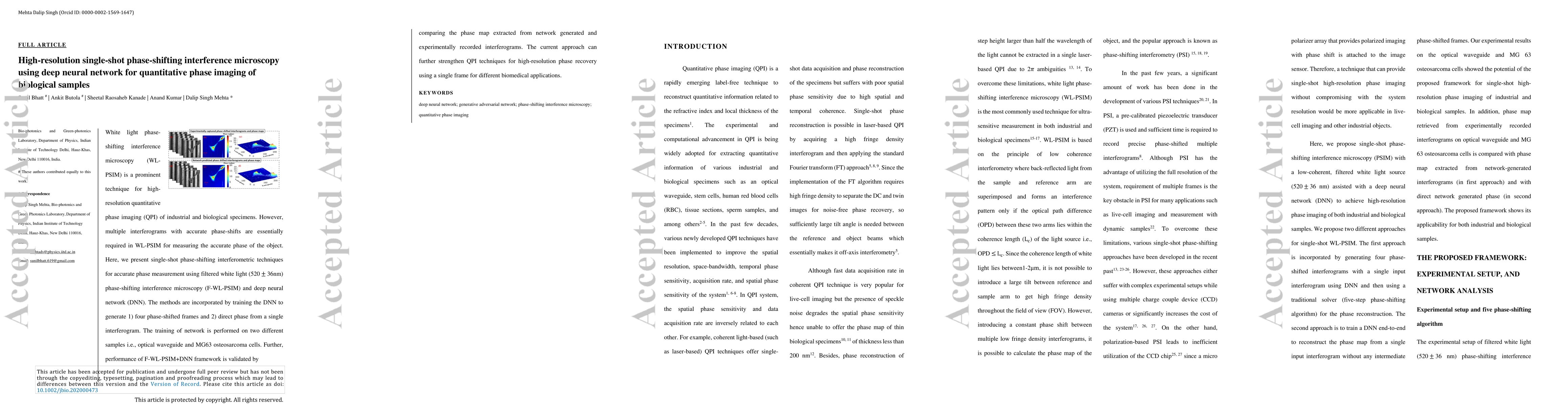
White light phase-shifting interference microscopy (WL-PSIM) is a prominent technique for high-resolution quantitative phase imaging (QPI) of industrial and biological specimens. However, multiple interferograms with accurate phase-shifts are essentially required in WL-PSIM for measuring the accurate phase of the object. Here, we present single-shot phase-shifting interferometric techniques for accurate phase measurement using filtered white light phase-shifting interference microscopy (F-WL-PSIM) and deep neural network (DNN). The methods are incorporated by training the DNN to generate 1) four phase-shifted frames and 2) direct phase from a single interferogram. The training of network is performed on two different samples i.e., optical waveguide and MG63 osteosarcoma cells. Further, performance of F-WL-PSIM+DNN framework is validated by comparing the phase map extracted from network generated and experimentally recorded interferograms. The current approach can further strengthen QPI techniques for high-resolution phase recovery using a single frame for different biomedical applications.
AI Key Findings
Get AI-generated insights about this paper's methodology, results, significance, and more — seven facets brought into focus.
Impact
Paper Details
PDF Preview
Key Terms
Citation Network
Current paper (gray), citations (green), references (blue)
Display is limited for performance on very large graphs.
Discussion 0