ArxivLens

Quick Summary:

This paper introduces a high-speed, label-free intensity diffraction tomography technique called annular illumination (aIDT) that rapidly characterizes large-volume 3D refractive index distributions in vitro. The method achieves 10 Hz volumetric rates with minimal data requirements, enabling detailed 3D quantitative phase imaging of complex biological samples.

DOI: 10.1117/1.AP.1.6.066004
arXiv Id: 1904.06004
Date Published: 2020-01-01
Date Updated: 2020-01-01

Key Terms (TF-IDF):

illumination (0.252)
technique (0.178)
high speed (0.175)
volume (0.161)
diffraction (0.161)
cellular (0.157)
label (0.148)
recover (0.144)
intensity (0.140)
https github (0.140)
github com (0.138)
resolution (0.138)
com (0.136)
mu (0.133)
https (0.132)
github (0.132)
speed (0.130)
imaging (0.128)
samples (0.127)
free (0.126)
License: http://arxiv.org/licenses/nonexclusive-distrib/1.0/

Summary:

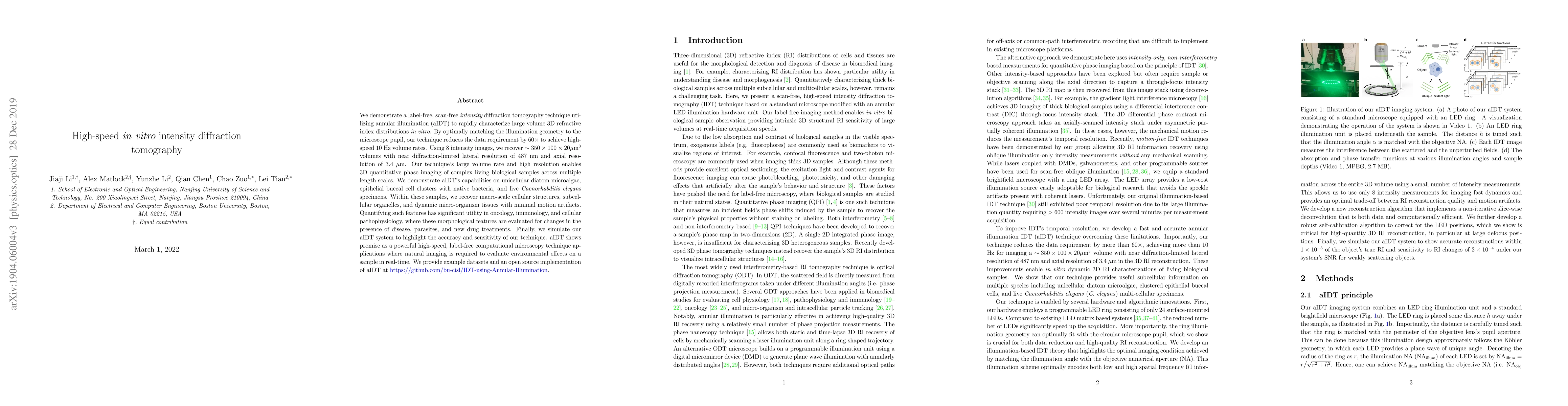
We demonstrate a label-free, scan-free {\it intensity} diffraction tomography technique utilizing annular illumination (aIDT) to rapidly characterize large-volume 3D refractive index distributions in vitro. By optimally matching the illumination geometry to the microscope pupil, our technique reduces the data requirement by 60$\times$ to achieve high-speed 10 Hz volume rates. Using 8 intensity images, we recover $\sim350\times100\times20\mu$m$^3$ volumes with near diffraction-limited lateral resolution of 487 nm and axial resolution of 3.4 $\mu$m. Our technique's large volume rate and high resolution enables 3D quantitative phase imaging of complex living biological samples across multiple length scales. We demonstrate aIDT's capabilities on unicellular diatom microalgae, epithelial buccal cell clusters with native bacteria, and live \emph{Caenorhabditis elegans} specimens. Within these samples, we recover macro-scale cellular structures, subcellular organelles, and dynamic micro-organism tissues with minimal motion artifacts. Quantifying such features has significant utility in oncology, immunology, and cellular pathophysiology, where these morphological features are evaluated for changes in the presence of disease, parasites, and new drug treatments. Finally, we simulate our aIDT system to highlight the accuracy and sensitivity of our technique. aIDT shows promise as a powerful high-speed, label-free computational microscopy technique applications where natural imaging is required to evaluate environmental effects on a sample in real-time. We provide example datasets and an open source implementation of aIDT at \href{https://github.com/bu-cisl/IDT-using-Annular-Illumination}{https://github.com/bu-cisl/IDT-using-Annular-Illumination}.

Citation & Reference Network Visualization

This graph shows how this paper is connected to other research. The current paper is shown in dark gray, papers that cite it are in green, and papers it references are in blue. You can:

Current Paper
Papers citing this work (Citations)
Papers cited by this work (References)

• Hover over any node to see the paper title

• Click a node to view that paper's details

• Drag nodes to rearrange the graph

• Use the zoom controls (bottom right) to zoom in/out

Citations and References

Title Authors Year Actions
Tailored 3D microphantoms: An essential tool for quantitative phase tomography analysis of organoids
M. Ziemczonok, S. Desissaire, Jeremy Neri, A. Kuś, L. Hervé, C. Fiche, G. Godefroy, Marie Fackeure, Damien Sery, W. Krauze, Kiran Padmanabhan, C. Paviolo, Małgorzata Kujawińska
2024
View Paper
Three-dimensional refractive index microscopy based on the multi-layer propagation model with obliquity factor correction
Zhan Tong, Xuesong Ren, Zihan Zhang, Boyang Wang, Yubin Miao, Guoxiang Meng
2024
View Paper
Recent Advances and Current Trends in Transmission Tomographic Diffraction Microscopy
N. Verrier, M. Debailleul, O. Haeberlé
2024
View Paper
AI-driven projection tomography with multicore fibre-optic cell rotation
Jiawei Sun, Bin Yang, N. Koukourakis, Jochen Guck, Juergen W. Czarske
2023
View Paper
Micro-tomographic and infrared spectral data mining for breast cancer diagnosis
Shan-yu Guo, J. Xiu, Lingnan Kong, Xingyu Kong, Hanqiu Wang, Zhiwei Lü, Famei Xu, Jing Li, Te Ji, Fuli Wang, Huiqiang Liu
2023
View Paper
Live-cell analysis framework for quantitative phase imaging with slightly off-axis digital holographic microscopy
Qian Shen, Zhuoshi Li, Jiasong Sun, Yao Fan, Yuanyuan Chen, Haojie Gu, P. Gao, Qian Chen, Chao Zuo
2022
View Paper
Speckle suppression using F-D2NN in holographic display
Jun Wang, Huang Yang, Yang Wu, Fan Wang
2022
View Paper
Extended coherent modulation imaging for single-shot object retrieval free from illumination artifacts.
Jiangtao Zhao, Wenhui Xu, Jianji Yi, Bingyang Wang, Fucai Zhang
2022
View Paper
Asymmetrical Illumination Enables Lipid Droplets Segmentation in Caenorhabditis elegans Using Epi-Illumination Dark Field Microscopy
Ruijie Shi, Yu Sun, J. Fang, Xiangyang Chen, Zachary J Smith, K. Chu
2022
View Paper
Tensorial tomographic differential phase-contrast microscopy
Shiqi Xu, Xiang Dai, Xi Yang, Kevin C Zhou, Kanghyun Kim, Vinayak Pathak, C. Glass, R. Horstmeyer
2022
View Paper
Optical diffraction tomography meets metrology—Measurement accuracy on cellular and subcellular level
M. Ziemczonok, A. Kuś, M. Kujawińska
2022
View Paper
Partially Coherent Illumination Based Point-Diffraction Digital Holographic Microscopy Study Dynamics of Live Cells
Kequn Zhuo, Yu Wang, Yang Wang, Kai Wen, Min Liu, Ying Ma, Juanjuan Zheng, P. Gao
2021
View Paper
Structured illumination microscopy with partially coherent illumination for phase and fluorescent imaging.
Kai Wen, Zhao-zhao Gao, X. Fang, Min Liu, Juanjuan Zheng, Ying Ma, Z. Zalevsky, P. Gao
2021
View Paper
Optimization analysis of partially coherent illumination for refractive index tomographic microscopy
Jiaji Li, Ning Zhou, Zhidong Bai, Shun Zhou, Qian Chen, Chao Zuo
2021
View Paper
Common-path intrinsically achromatic optical diffraction tomography.
P. Zdańkowski, J. Winnik, K. Patorski, P. Gocłowski, M. Ziemczonok, M. Józwik, M. Kujawińska, Maciej Trusiak
2021
View Paper
Partially Coherent Optical Diffraction Tomography Toward Practical Cell Study
J. Soto, J. Rodrigo, T. Alieva
2021
View Paper
Smart computational light microscopes (SCLMs) of smart computational imaging laboratory (SCILab)
Yao Fan, Jiaji Li, Linpeng Lu, Jiasong Sun, Yan Hu, Jialin Zhang, Zhuoshi Li, Qian Shen, Bowen Wang, Runnan Zhang, Qian Chen, Chao Zuo
2021
View Paper
Snapshot Partially Coherent Diffraction Tomography
Bo Xiong, Xiaoxu Li, You Zhou, Liejun Wang, Jiamin Wu, Qionghai Dai
2021
View Paper
Optimizing sample illumination scanning in transmission tomographic diffractive microscopy.
A. Taddese, N. Verrier, M. Debailleul, Jean-Baptiste Courbot, O. Haeberlé
2021
View Paper
Optronic convolutional neural networks of multi-layers with different functions executed in optics for image classification.
Ziyu Gu, Yesheng Gao, Xingzhao Liu
2021
View Paper
Concept, implementations and applications of Fourier ptychography
G. Zheng, Cheng Shen, Shaowei Jiang, Pengming Song, Changhuei Yang
2021
View Paper
Concept, implementations and applications of Fourier ptychography
G. Zheng, Cheng Shen, Shaowei Jiang, Pengming Song, Changhuei Yang
2021
View Paper
Spatial resolution in photoacoustic computed tomography
Chao Tian, Chenxi Zhang, H. Zhang, D. Xie, Yi Jin
2021
View Paper
Transport of intensity equation: a tutorial
Chao Zuo, Jiaji Li, Jiasong Sun, Fan Yao, Jialin Zhang, Linpeng Lu, Runnan Zhang, Bowen Wang, Lei Huang, Qian Chen
2020
View Paper
A versatile and customizable low-cost 3D-printed open standard for microscopic imaging
Benedict Diederich, René Lachmann, S. Carlstedt, Barbora Marsíková, Haoran Wang, Xavier Uwurukundo, A. Mosig, R. Heintzmann
2020
View Paper
Resolution-enhanced intensity diffraction tomography in high numerical aperture label-free microscopy
Jiaji Li, Alex Matlock, Yunzhe Li, Qian Chen, L. Tian, Chao Zuo
2020
View Paper
High-performance imaging of cell-substrate contacts using refractive index quantification microscopy.
Ziqiang Xin, Chonglei Zhang, Lixun Sun, Chao Wan, Ting Chen, Houkai Chen, Min Wang, Yijia Wang, Siwei Zhu, Xiaocong Yuan
2020
View Paper
Label-free quantitative 3D intensity diffraction tomographic imaging in high numerical aperture microscopy
Jiaji Li, Alex Matlock, Yunzhe Li, Qian Chen, L. Tian, Chao Zuo
2020
View Paper
Optimization method for the synthetic apertures imaging system
Bowen Wang, Yan Zou, Hao Xu, Yan Hu, Chao Zuo
2020
View Paper
Holographic lensless quantitative phase imaging microscope
Linpeng Lu, Xiangyu Zhang, Jiasong Sun, Jialin Zhang, Yao Fan, Qian Chen, Chao Zuo
2020
View Paper
Miniaturized multi-contrast quantitative phase imaging microscope
Yao Fan, Junyi Ding, Jiasong Sun, Qian Chen, Chao Zuo
2020
View Paper
Physics-embedded deep learning for intensity diffraction tomography
Alex Matlock, L. Tian
2020
View Paper
Fast compressive lens-free tomography for 3D biological cell culture imaging.
Zhenxiang Luo, A. Yurt, R. Stahl, M. Carlon, A. Ramalho, F. Vermeulen, A. Lambrechts, D. Braeken, L. Lagae
2020
View Paper
High-resolution and large field-of-view Fourier ptychographic microscopy and its applications in biomedicine
An Pan, Chao Zuo, Baoli Yao
2020
View Paper
UC2 – A Versatile and Customizable low-cost 3D-printed Optical Open-Standard for microscopic imaging
Benedict Diederich, René Lachmann, S. Carlstedt, Barbora Marsíková, Haoran Wang, Xavier Uwurukundo, A. Mosig, R. Heintzmann
2020
View Paper
Inverse scattering for reflection intensity phase microscopy.
Alex Matlock, A. Sentenac, P. Chaumet, Ji Yi, L. Tian
2019
View Paper
Diffraction tomography with a deep image prior.
Kevin C Zhou, R. Horstmeyer
2019
View Paper
SIMBA: Scalable Inversion in Optical Tomography Using Deep Denoising Priors
Zihui Wu, Yu Sun, Alex Matlock, Jiaming Liu, L. Tian, U. Kamilov
2019
View Paper
High-speed in vitro intensity diffraction tomography
Jiaji Li, Alex Matlock, Yunzhe Li, Qian Chen, Chao Zuo, L. Tian
2019
View Paper
Wide-field high-resolution 3D microscopy with Fourier ptychographic diffraction tomography
Chao Zuo, Jiasong Sun, Jiaji Li, A. Asundi, Qian Chen
2019
View Paper
MPIS-IDTM: Multi-plane assisted iterative solution against scattering in intensity diffraction tomographic microscopy
Jiaji Li, Wenjun Shao, Tianliang Li, Xingang Mou
2025
View Paper
Imaging through weak scattering media via high-order Fourier ptychographic microscopy
Jinghao Xu, Hanbing Chen, Yizheng Liao, Tianci Feng, An Pan
2025
View Paper
Extended depth of field reconstruction with complex field estimation
Jing Hu, Sunzheng Li, Yibing Shen, Jinlei Zhang, Zhenrong Zheng
2022
View Paper
Absorption and phase decoupling in transport of intensity diffraction tomography
Zhidong Bai, Qian Chen, H. Ullah, Linpeng Lu, N. Zhou, Shun Zhou, Jiaji Li, Chao Zuo
2022
View Paper

Similar Papers

Bond-Selective Intensity Diffraction Tomography

Biao Wang, Yihong Xu, Hongbo Zhu, Jian Zhao, Lei Tian, Alex Matlock, Fukai Chen, Ji-Xin Cheng, Jiabei Zhu, Zhicong Chen, Ziqi Song, Yuewei Zhan, Xingchen Lin

Published: 2023-01-11

Comments (0)

This website uses cookies to ensure you get the best experience. Learn more