The study explores advanced fluorescence microscopy techniques, particularly 2P and 1P voltage imaging, combined with novel voltage indicators and imaging systems to capture fast neural dynamics with high spatiotemporal resolution.
High-speed optical microscopy for neural voltage imaging: Methods, trade-offs, and opportunities
Publication
Metrics
Quick Answers
What methodology did the authors use?
The study explores advanced fluorescence microscopy techniques, particularly 2P and 1P voltage imaging, combined with novel voltage indicators and imaging systems to capture fast neural dynamics with high spatiotemporal resolution. More in Methodology →
What are the key results?
2P voltage imaging enables deep brain imaging with high spatiotemporal resolution — Compressed sensing and ROI-matched data acquisition significantly reduce data volume while preserving essential information More in Key Results →
Why is this work significant?
This research advances the capability to study neural circuits and dynamics in vivo with unprecedented resolution, offering insights into brain function, behavior, and disease mechanisms. More in Significance →
What are the main limitations?
Current 2P voltage imaging is limited by phototoxicity and tissue heating at higher excitation powers — Large FOV voltage imaging remains constrained by technical challenges in photon collection and camera readout speed More in Limitations →
Abstract
High-speed optical imaging of dynamic neuronal activity is essential yet challenging in neuroscience. While calcium imaging has been firmly established as a workhorse technique for monitoring neuronal activity, its limited temporal resolution and indirect measurement restrict its ability to capture rapid inhibitory and excitatory events and subthreshold voltage oscillations. In contrast, voltage imaging directly measures membrane potential fluctuations, providing a comprehensive and precise representation of neuronal circuit dynamics. Recent advancements in voltage-sensitive dyes and, particularly, genetically encoded voltage indicators have significantly enhanced the feasibility of voltage imaging, prompting the development of advanced fluorescence microscopy methods optimized for high-speed acquisition. However, achieving millisecond-scale temporal resolution remains challenging due to inherent trade-offs among imaging speed, spatial resolution, and signal-to-noise ratio. Conventional raster-scanning approaches, including confocal microscopy, are fundamentally limited by their slow frame rates, precluding the capture of rapid neuronal events from multiple neurons simultaneously. Alternative techniques such as random-access scanning, spatiotemporal multiplexing, and computational optical imaging have successfully addressed these constraints, enabling kilohertz-level imaging of neuronal activity in both two-dimensional and three-dimensional contexts. This review summarizes recent progress in high-speed optical microscopy for voltage imaging and discusses its transformative potential for neuroscience research.
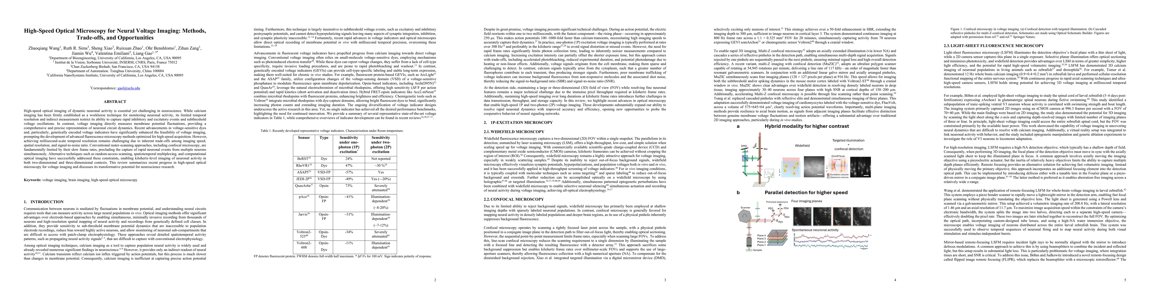
Key Findings, in focus
Seven facets of this paper, analysed and brought into focus by AI.
This research advances the capability to study neural circuits and dynamics in vivo with unprecedented resolution, offering insights into brain function, behavior, and disease mechanisms.
- 2P voltage imaging enables deep brain imaging with high spatiotemporal resolution
- Compressed sensing and ROI-matched data acquisition significantly reduce data volume while preserving essential information
- Miniaturized head-mounted systems allow for voltage imaging in freely moving animals
This research advances the capability to study neural circuits and dynamics in vivo with unprecedented resolution, offering insights into brain function, behavior, and disease mechanisms.
The work introduces novel imaging strategies, including scan multiplier units, spatio-temporal multiplexing, and adaptive excitation sources, to enhance imaging throughput and reduce phototoxicity.
This research combines cutting-edge voltage indicators with innovative imaging systems and data acquisition techniques, enabling previously unattainable insights into neural activity across diverse experimental conditions.
- Current 2P voltage imaging is limited by phototoxicity and tissue heating at higher excitation powers
- Large FOV voltage imaging remains constrained by technical challenges in photon collection and camera readout speed
- Development of brighter and more sensitive GEVIs for NIR imaging
- Integration of compressive sensing with advanced camera technologies for large-scale voltage imaging
- Optimization of spatiotemporal de-mixing algorithms for single-neuron resolution
Discussion 0