High-speed scattering polarimetry for correlative nerve fiber imaging and multi-modal analysis
Publication
Metrics
AI Quick Summary
This research introduces the Scattering Polarimeter, a high-speed microscope combining 3D Polarized Light Imaging (3D-PLI) and Computational Scattered Light Imaging (ComSLI) to map dense nerve fibers in brain sections. The device generates detailed multi-modal fiber direction maps, integrating precise fiber orientations from 3D-PLI with complex fiber crossings from ComSLI, achieving results comparable to existing setups.
Paper Preview
Abstract
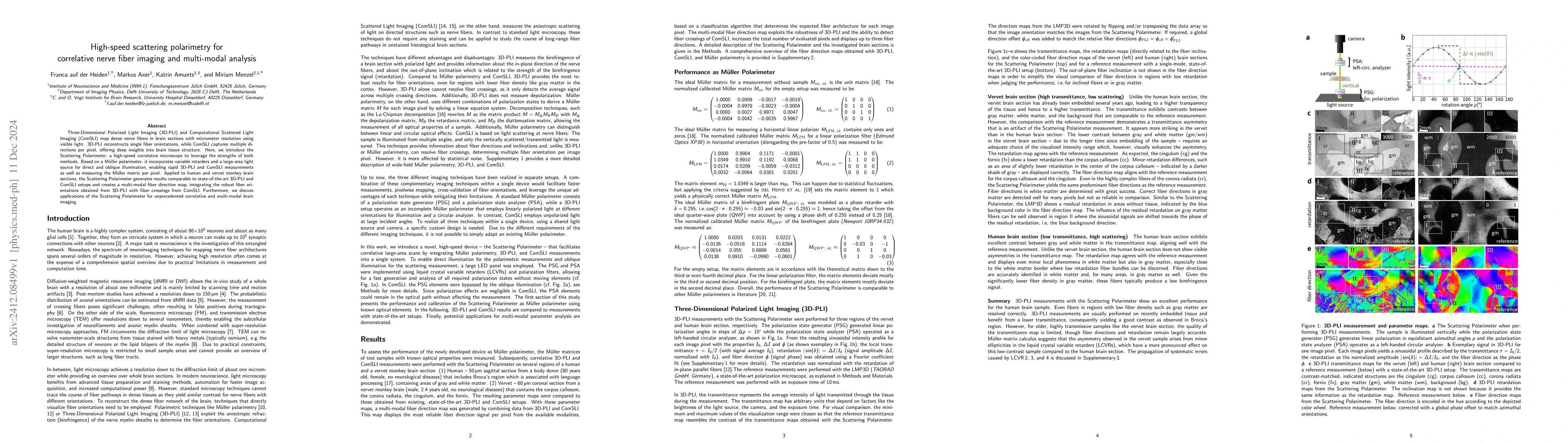
Three-Dimensional Polarized Light Imaging (3D-PLI) and Computational Scattered Light Imaging (ComSLI) map dense nerve fibers in brain sections with micrometer resolution using visible light. 3D-PLI reconstructs single fiber orientations, while ComSLI captures multiple directions per pixel, offering deep insights into brain tissue structure. Here, we introduce the Scattering Polarimeter, a high-speed correlative microscope to leverage the strengths of both methods. Based on a M\"uller polarimeter, it incorporates variable retarders and a large-area light source for direct and oblique illumination, enabling rapid 3D-PLI and ComSLI measurements as well as measuring the M\"uller matrix per pixel. Applied to human and vervet monkey brain sections, the Scattering Polarimeter generates results comparable to state-of-the-art 3D-PLI and ComSLI setups and creates a multi-modal fiber direction map, integrating the robust fiber orientations obtained from 3D-PLI with fiber crossings from ComSLI. Furthermore, we discuss applications of the Scattering Polarimeter for unprecedented correlative and multi-modal brain imaging.
AI Key Findings
Get AI-generated insights about this paper's methodology, results, significance, and more — seven facets brought into focus.
Paper Details
Authors
PDF Preview
Related Papers
No references found for this paper.
Discussion 0