HistomicsML2.0: Fast interactive machine learning for whole slide imaging data
Publication
Metrics
AI Quick Summary
A new software tool called HistomicsML2.0 enables rapid training of machine learning classifiers for detecting histologic patterns in whole-slide images, making it easier for non-experts to analyze medical data.
Paper Preview
Abstract
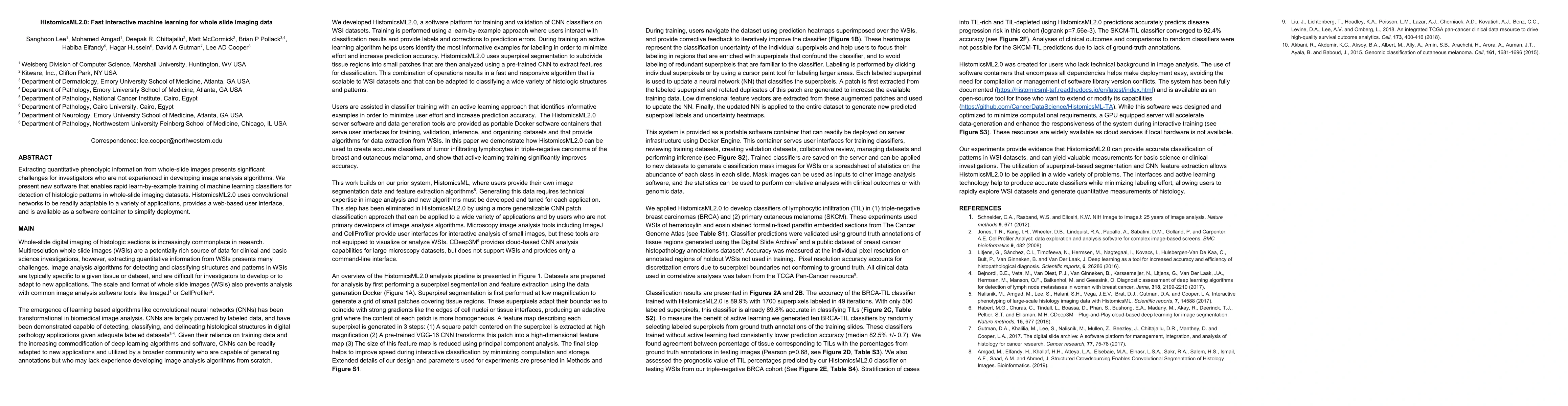
Extracting quantitative phenotypic information from whole-slide images presents significant challenges for investigators who are not experienced in developing image analysis algorithms. We present new software that enables rapid learn-by-example training of machine learning classifiers for detection of histologic patterns in whole-slide imaging datasets. HistomicsML2.0 uses convolutional networks to be readily adaptable to a variety of applications, provides a web-based user interface, and is available as a software container to simplify deployment.
AI Key Findings
Get AI-generated insights about this paper's methodology, results, significance, and more — seven facets brought into focus.
Impact
Paper Details
Authors
PDF Preview
Key Terms
Citation Network
Current paper (gray), citations (green), references (blue)
Display is limited for performance on very large graphs.
Discussion 0